We have another new series of posts to offer this month- Point of Care Ultrasound in Nephrology (POCUN). What better way start off than with kidney stones.
Urolithiasis is one of the most common indications for urinary tract ultrasonography – whether you are dealing with a patient with flank pain, looking for source of obstruction in hydronephrosis, or following up on stone burden in your clinic patient.
In general, something that reflects most of the ultrasound waves back (or makes more echoes) will appear bright or hyperechoic. For example, stones, bone, fibrous tissue ( diaphragm, renal capsule) and adipose tissue (renal sinus fat) are some of the bright structures seen on abdominal ultrasonography images.
There are two useful artifacts that will help us identify stones and calcified structures: acoustic shadowing and twinkle artifact.
Acoustic Shadowing
Acoustic shadowing is the black (anechoic) or hypoechoic band seen beyond echogenic structures that do not transmit ultrasound waves such as stone and bone. It is similar to us forming a shadow when we are in the pathway of light. Fat and fibrous structures allow some sound waves to pass through them and therefore typically do not give shadowing. However, depending exclusively on shadowing to diagnose stones may lead to false negative interpretation (especially with small stones). When a stone or calcification is not large enough to block the cross section of the ultrasound beam, shadowing will not occur. Also, shadowing may not be apparent in poor quality pictures with overall increased gain. The twinkle artifact seen on the color Doppler can be of great help in these situations
Twinkle Artifact
In the color Doppler mode, the velocity of mobile acoustic interfaces such as red blood cells is measured as a shift in frequency and represented as a range of colors: red color denotes flow towards the transducer and blue away from the transducer. A mosaic pattern of colors is seen in regions of turbulent flow. Stones demonstrate twinkling in this mode, which is a rapidly alternating focus of intense color signals resembling turbulent flow that is more pronounced with rougher stones. It appears with or without an associated color comet tail artifact. Twinkling can be seen just on the stone or following like a band or tail past it. One thing to remember is that if you set the focal zone below the stone, twinkling will be more pronounced compared to setting it above. Focal zone is a user controlled feature on the machine which gives best definition at a designated depth of focus – it’s like tapping to change the camera focus on your smartphone. Of note, the sensitivity and specificity of twinkle artifact for recognition of stones less than 5 mm were 99.1% and 90.9% respectively in one study.
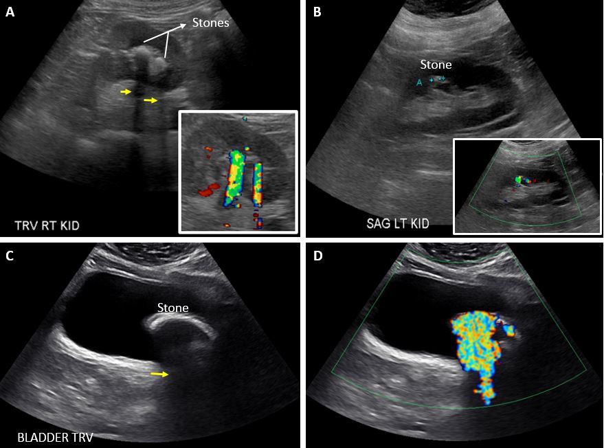
Figure A shows transverse view of the kidney with two stones appearing as echogenic foci accompanied by shadowing (arrows). Corresponding twinkle artifact with comet tail is shown in the inset. Figure B shows a stone without prominent shadowing but with twinkling. Note this twinkle doesn’t have a tail. Figure C demonstrates a bladder stone with shadow (arrow) and corresponding twinkle artifact with comet tail can be seen in Figure D.
A recommendation: obtain a few Doppler images when doing a kidney ultrasound!
Post by: Abhilash Koratala, MD, FASN (@NephroP)
University of Florida


thanks for i really love this website
Best solution for kidney patients, thank you
Good treatment for kidney patients, thank you
Thanks for sharing this information.
Very nice.
Amazing tricks