Anti-Nephrin Antibodies in Minimal Change Disease
Approach to the Patient with Peritoneal Dialysis Catheter Dysfunction- Part 1
Want to write a case report? RFN has you covered with a list of journals to choose from
Have a Nephrology Question? There Might Be A #Tweetorial For That!
Conferences, Courses, & Grants
FOAMed
Interventional Nephrology Series
Focus on Point-of-Care Ultrasound in Nephrology (POCUN) Series
Kidney Biopsy of the Month Series
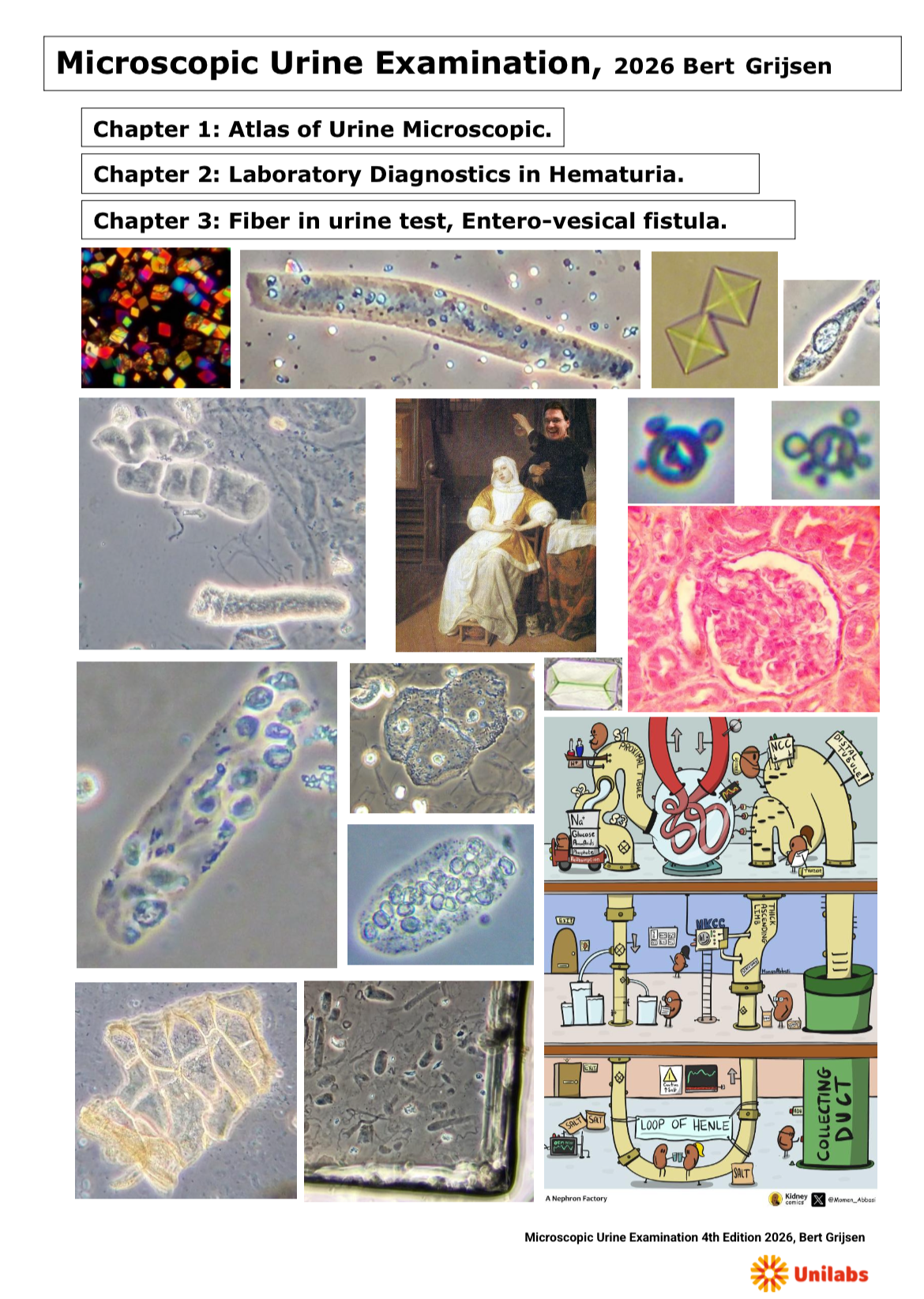
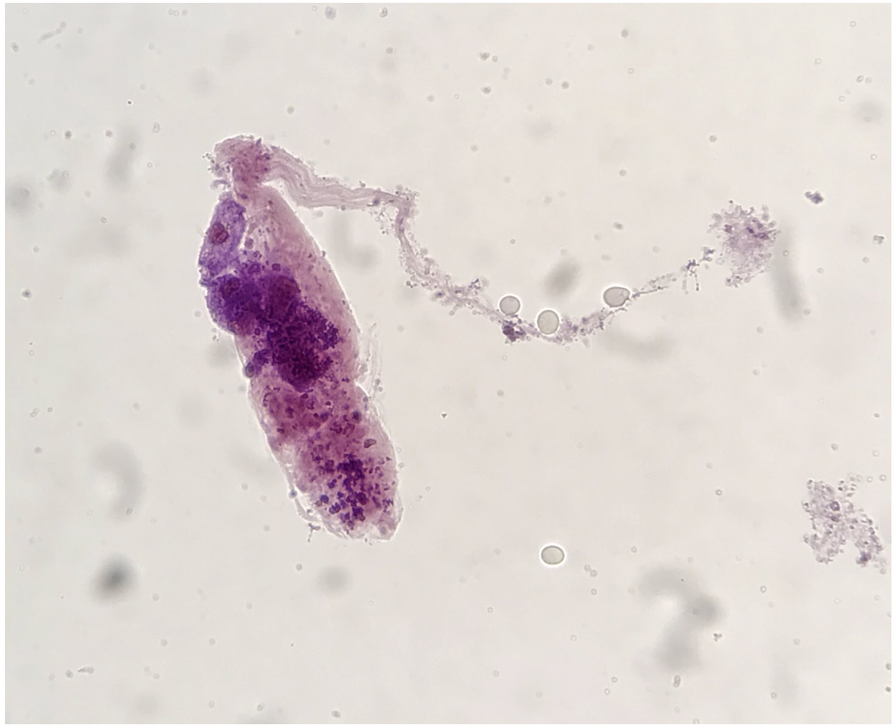
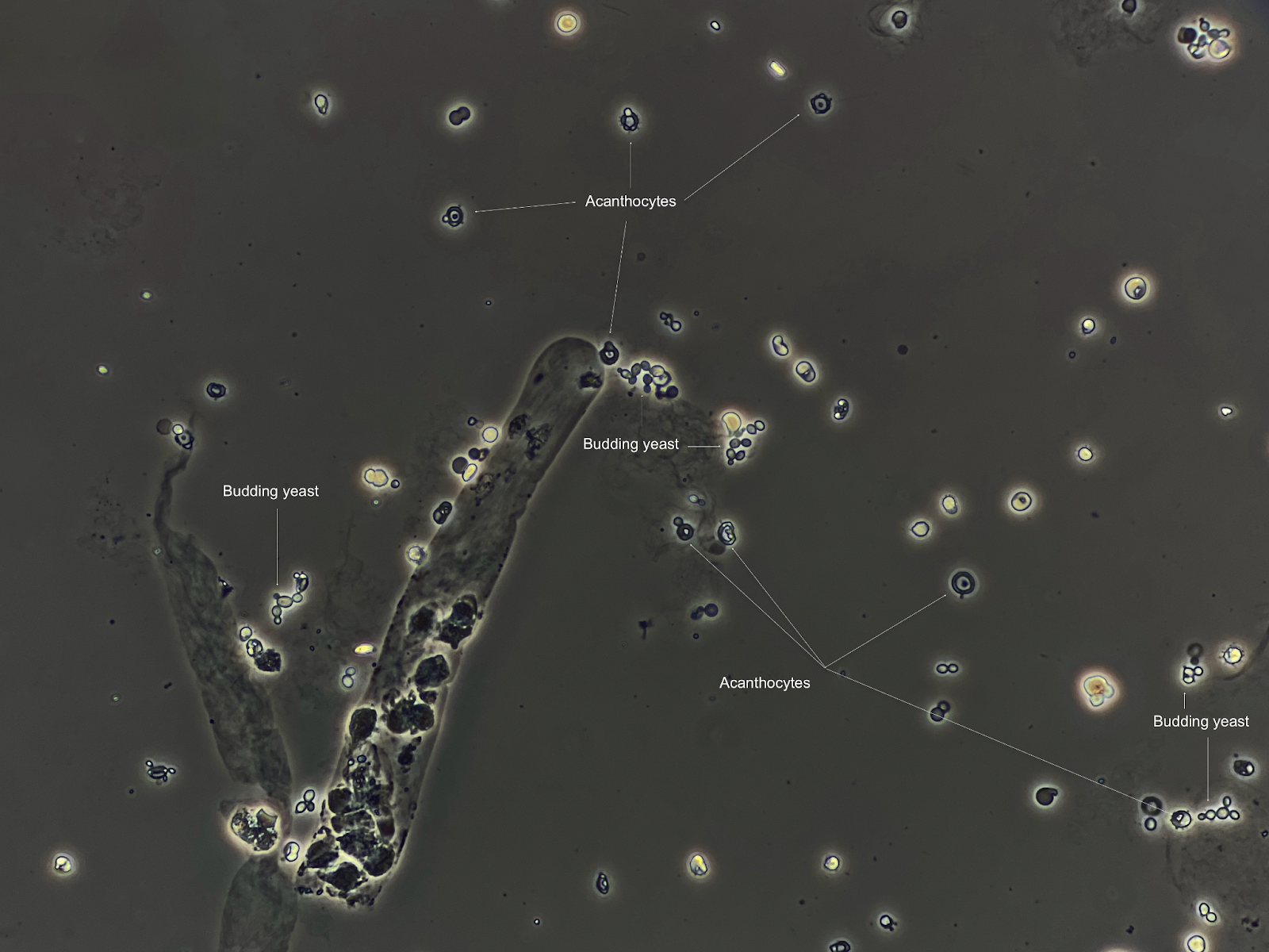
Microscopic Urine Sediment Examination Atlas
This “Atlas”: Microscopic Urine Examination compiled in 2026 by Bert Grijsen, is freely accessible to everyone, meets the needs of those interested in correctly performing and evaluating microscopic urine examinations. It contains microscopic images of the most common…
Leucine Crystals in the Urine – Still enigmatic after more than 150 years
Exemplary case A 65-year-old patient with a history of cholangiocarcinoma was admitted with abdominal pain, increasing ascites and jaundice. He was diagnosed with spontaneous bacterial peritonitis. Shortly afterwards he developed septic shock and an acute kidney injury. The…
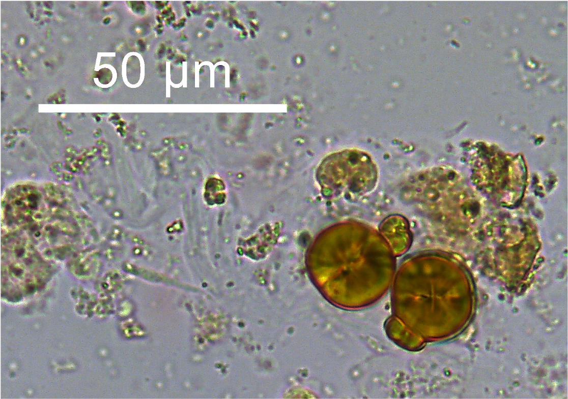
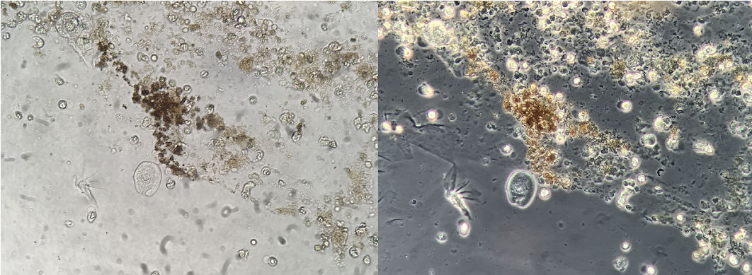
Cylindroids in Urine, Have You Ever Seen It?
The German Louis G.F. Thomas (1838 —1907) described “cylindroids” (Figure 1) in 1870 to indicate elongated elements partly similar to casts [1], the nature of which is still unclear at present . There is no consensus on the…

Urine Sediment of the Month: Fibrin Casts in Urine Microscopy
Casts form in the distal tubules and collecting ducts when either Tamm-Horsfall mucoprotein or fibrin agglutinate and become molded into cylindrical structures within the tubular lumen. Cells and various substances, when present in the tubule, are incorporated into…
AKI in a patient with macroscopic hematuria
A 56-year-old kidney transplant recipient due to IgA nephropathy presented to the emergency department with a two day history of fatigue, gross hematuria, and pain over her allograft. She was diagnosed with transplant pyelonephritis and treated with antibiotics…
Urine Sediment of the Month: Another Look at Macroscopic Evaluation
Routine work with urine microscopy can lead to plenty of “surprises.” Sometimes not only the microscopic evaluation presents something new and unique, but the findings can be observed directly by looking at the sample. The original post on…
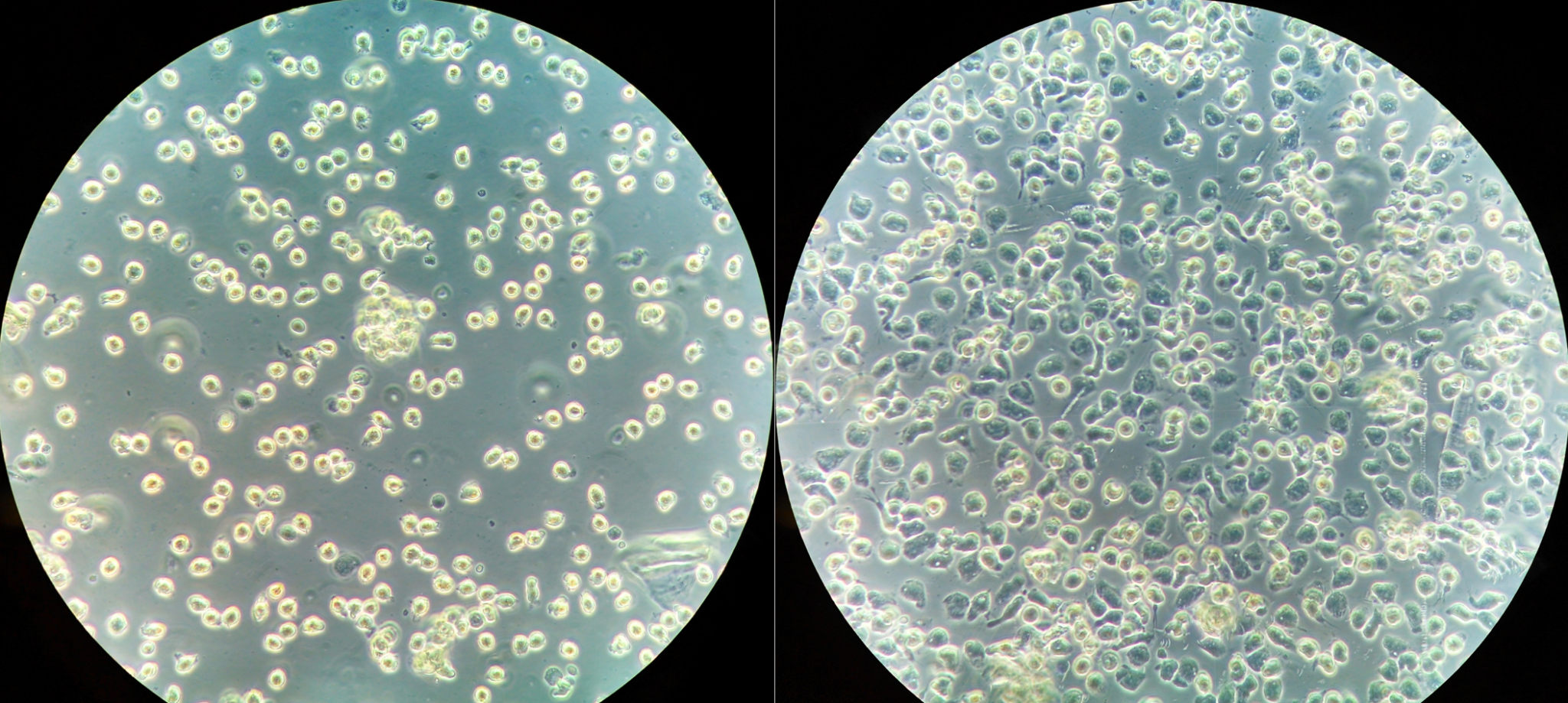
Urine Sediment of the Month: RBC “Lookalikes”
Red blood cells (RBCs) in the urine are normally 7-8 µm in diameter, circular or slightly oval and biconcave in appearance. They may appear crenated, with symmetric spiked membrane contour in hypertonic urine, or spherical, with near loss…
Leukocyte Lysis in Dilute Urine
The influence of urine concentration on the cells we observe during urine microscopy is well known. Concentrated urine specimens with a high urine specific gravity (SG) preserve nucleated cellular morphology the best, however, some cellular structures (e.g.: erythrocytes)…

Urine Sediment of the Month: The Thin Waxy Cast
The thickness of the fluid layer examined by urinary microscopy is determined by the volume of resuspended sediment put on the glass slide and the size of the coverslip. Using 13-15ul of sediment and coverslips of 18x18mm2 as…

The Case of the Missing Casts
A 50 y/o woman presents with symptoms of fatigue and malaise for 1-2 weeks. Medications include ACE, HCTZ, and a multivitamin daily. She had a UTI 6 weeks ago that did not improve after a 5 day course…