Casts form in the distal tubules and collecting ducts when either Tamm-Horsfall mucoprotein or fibrin agglutinate and become molded into cylindrical structures within the tubular lumen. Cells and various substances, when present in the tubule, are incorporated into these casts as they form, thus preserving evidence of what was going on in the tubules and collecting ducts at the time of cast formation. This information is often helpful in clarifying the nature of an underlying renal problem.
Tamm-Horsfall mucoprotein is secreted by cells lining the distal nephron and under certain conditions it agglutinates forming hyaline casts. The hyaline protein matrix is readily stained with Sternheimer-Malbin (SM) stain resulting in a pink or magenta color. Cells within this type of cast matrix are also subject to staining, with the degree of stain uptake related to cell viability (viable cells are able to resist stain uptake whereas older, non-viable, cells appear more stained.
When glomerular capillary rupture occurs, direct entry of blood into the tubular lumen leads to fibrin cast matrix formation. The fibrin protein matrix is typically not stained with SM stain and instead appears tan or yellow in color with a fibrous texture. Red blood cells within this matrix often appear weakly stained or unstained. Perhaps, casts with this appearance represent very ‘fresh’ casts and since the RBC’s are viable they are able to avoid stain uptake.
Further study is necessary to determine whether the presence of casts with a fibrin matrix correlates with the presence of glomerular capillary rupture and subsequent crescent formation on biopsy.
Here are examples of RBC and mixed cellular casts with “hyaline” and “fibrin” protein matrix:
Pictures A and B – RBC casts with a hyaline matrix (brightfield with SM stain), note the homogeneous stain uptake in the hyaline cast matrix and in the RBC’s.
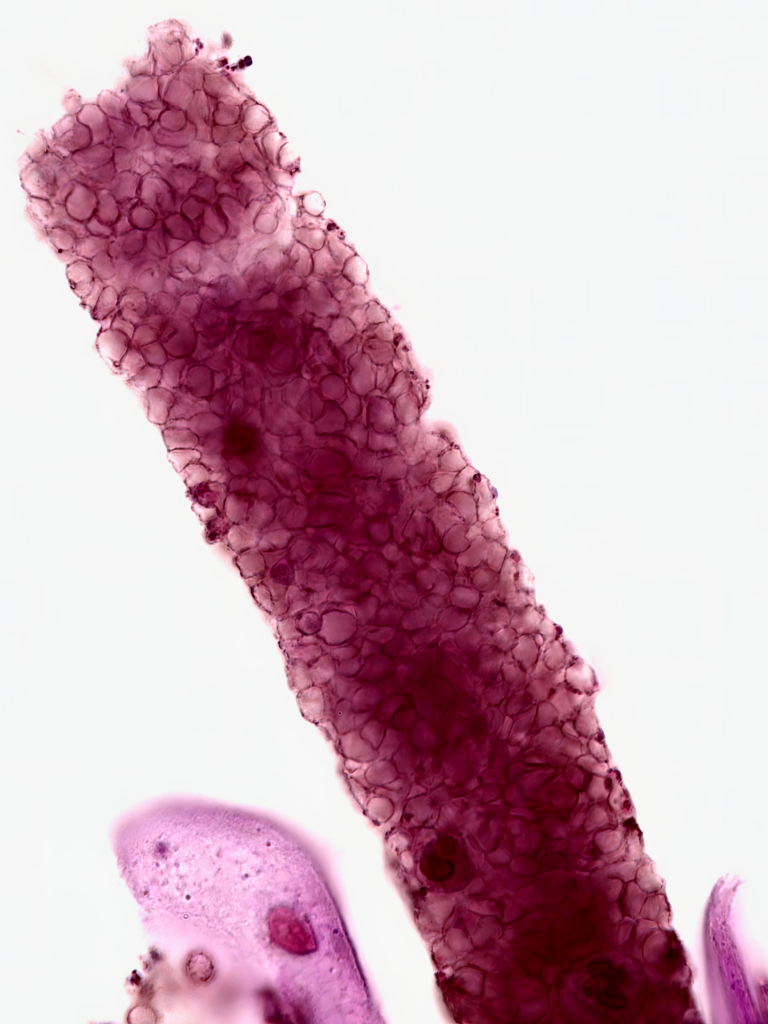
A
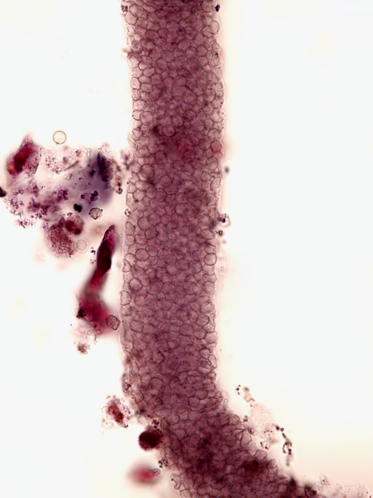
B
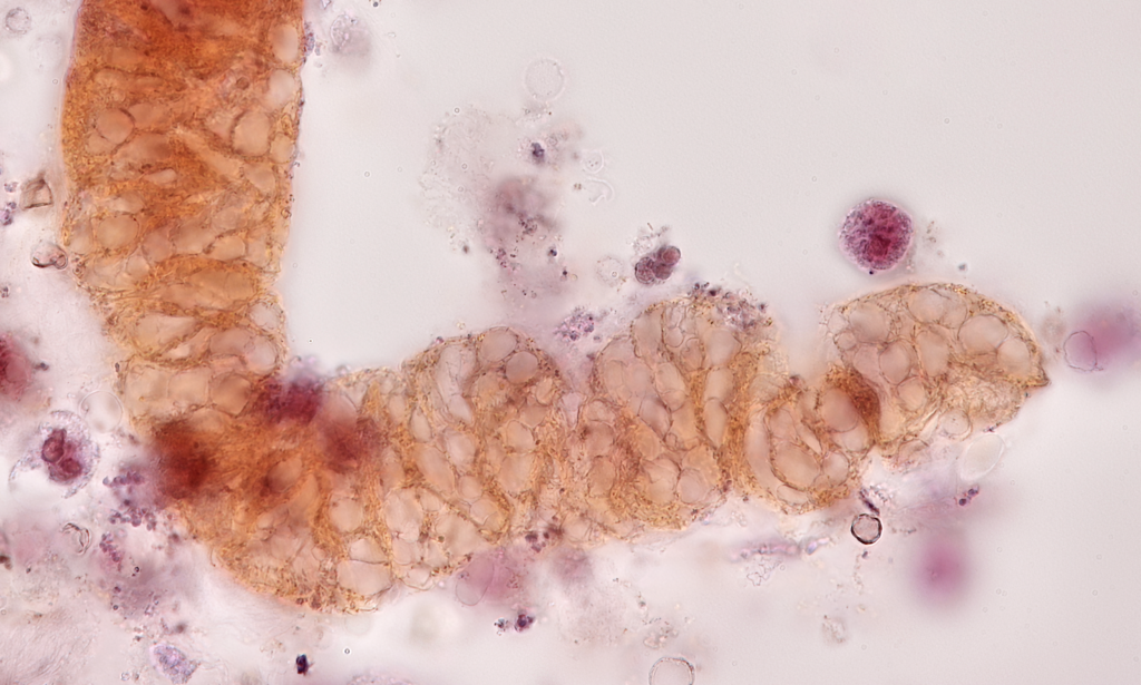
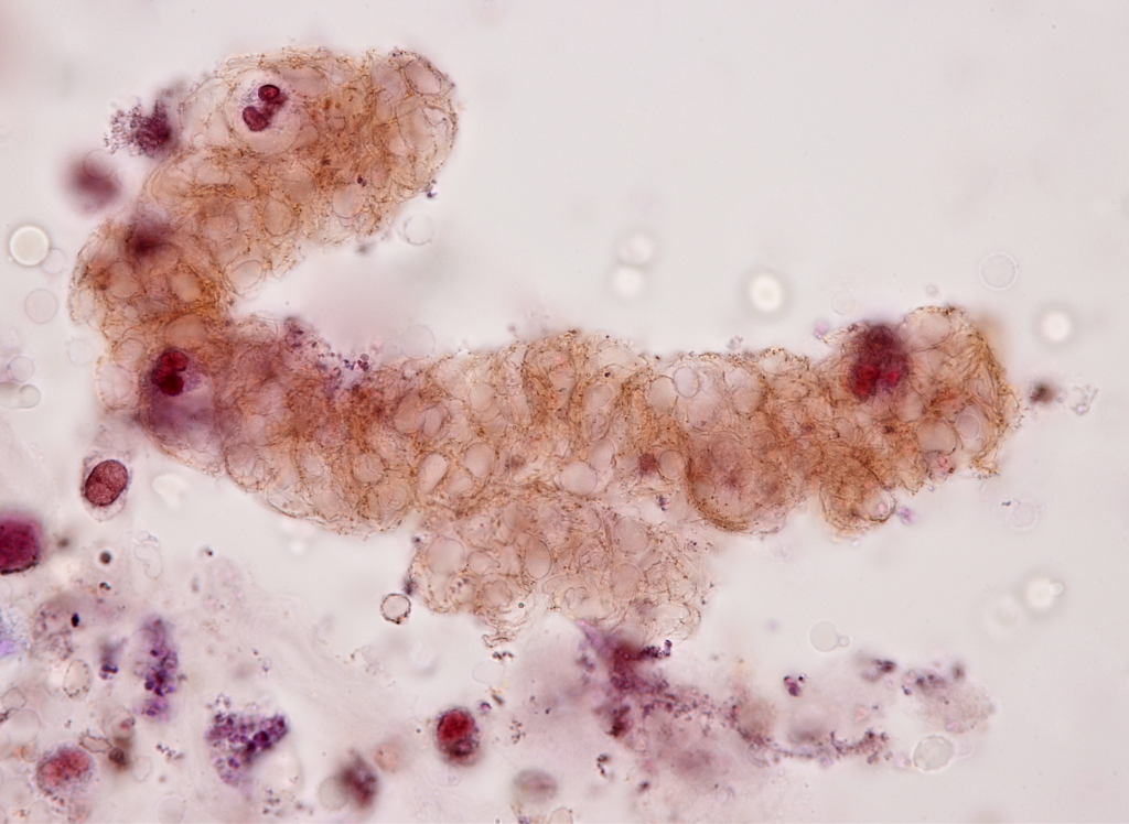
Pictures C,D,E, and F – RBC casts with a fibrin matrix (brightfield with SM stain), note the lack of stain uptake in the fibrin cast matrix and in the RBC’s. Also note fibrous character of protein matrix surrounding RBC’s
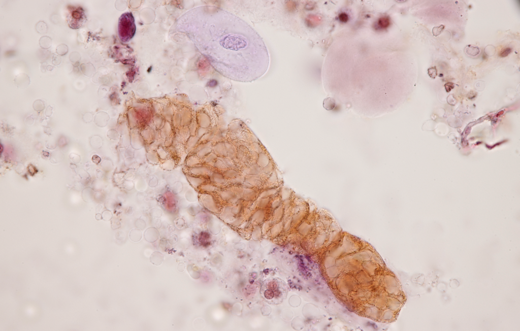
C
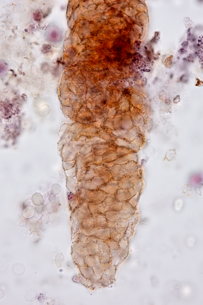
D
E
F
Pictures G and H – Mixed cellular casts, with RBC’s and a few WBC’s – brightfield with SM stain. Note lack of stain uptake in fibrin cast matrix and in the RBC’s. The WBC’s however appear to readily stain
G

H
By: Jay Seltzer
Reviewed by: Margaret DeOliveira

