Anti-Nephrin Antibodies in Minimal Change Disease
Approach to the Patient with Peritoneal Dialysis Catheter Dysfunction- Part 1
Want to write a case report? RFN has you covered with a list of journals to choose from
Have a Nephrology Question? There Might Be A #Tweetorial For That!
Conferences, Courses, & Grants
FOAMed
Interventional Nephrology Series
Focus on Point-of-Care Ultrasound in Nephrology (POCUN) Series
Kidney Biopsy of the Month Series

Pericardial effusion detected on point of care ultrasound: What next?
Abhilash Koratala, MD Medical College of Wisconsin In previous posts, we explored the identification of pericardial effusion through different cardiac windows using POCUS. Now that you know how to identify the effusion, let’s delve into the key findings…

POCUS orientation videos
Abhilash Koratala, MDMedical College of Wisconsin Learning POCUS is like acquiring physical examination skills. It takes time and involves gaining proficiency in image acquisition, interpretation, and clinical integration. While hands-on practice is a vital component, the learner must…
Identification of various effusions on standard echocardiographic views: part II
Post by: Abhilash Koratala, MD, FASN @NephroPMedical College of Wisconsin In the previous post, we discussed about the appearance of pericardial and pleural effusions in the parasternal long axis view. As parasternal short axis view is obtained from…

Point of Care Ultrasound for Nephrologists: Establishing Use Cases and Building the Evidence for Tomorrow
Check out an entire issue of Advances in Chronic Kidney Disease devoted to “Point-of-Care Ultrasound.” This issue was inspired by the growing body of young nephrologists who have become interested in using point-of-care ultrasound (POCUS) to improve outcomes…
Identification of Various Effusions on Standard Echocardiographic Views: Part 1
Abhilash Koratala, MD, FASN @NephroP, Medical College of Wisconsin Identification of pericardial effusion is one of the basic applications of focused cardiac ultrasound (FoCUS). As previously discussed, pericardial effusion appears as an anechoic or echo-free space between the…
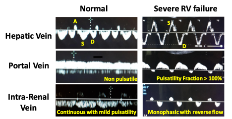
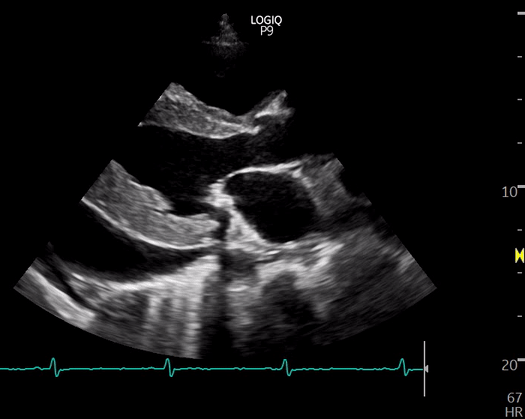
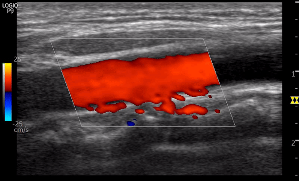
Evaluation of Venous Congestion Using Point of Care Ultrasonography
Eduardo R Argaiz @ArgaizR, National Institute of Medical Science and Nutrition Salvador Zubiran, Mexico It is well known that congestion is the primary reason for hospitalization in patients with acute heart failure and an important target for therapy….
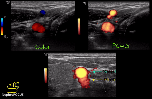
Basics of Doppler Ultrasound for the Nephrologist- Part 2
In the previous post, we talked about the Doppler effect, color flow and power Doppler modes. Now let us focus on the spectral Doppler. Spectral Doppler enables us to measure the velocity of blood flow as well as…
Basics of Doppler Ultrasound for the Nephrologist: Part 1
The greyscale ultrasound image is derived from the amplitude information in the echoes returning from a structure/organ. Rapidly moving structures such as red blood cells within blood vessels make very low amplitude echoes that cannot be displayed. That…
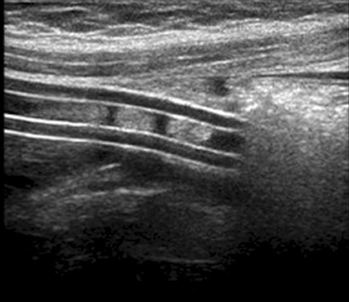
Bedside Ultrasonography of the Peritoneal Dialysis Catheter
Peritoneal dialysis (PD) catheters are one of the most successful long-term transcutaneous access devices ever used in medical practice. The catheter, if successfully placed can provide successful dialysis for years. For the above to be accomplished, various components…
An Overview of Lung Ultrasound: The COVID edition part 2
Last week we began with an introduction to lung ultrasound to equip us during the times of COVID-19. This week we dive into specific nuances that will aid our assessment of patients with COVID. Lung ultrasound for COVID-19…