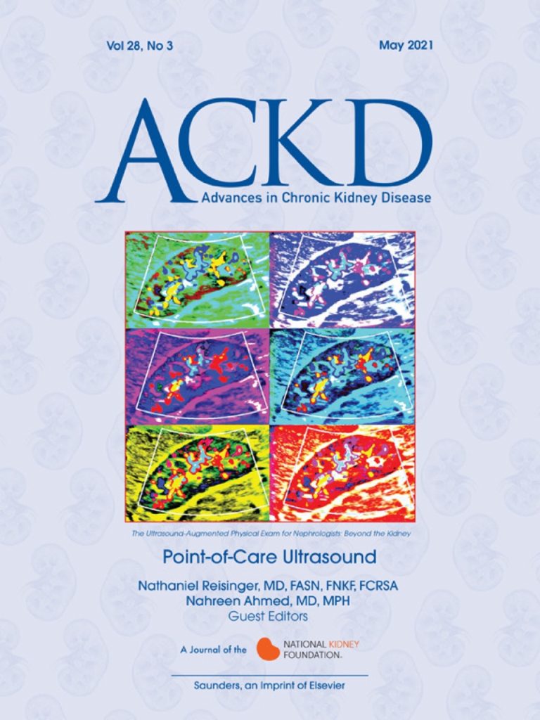
Check out an entire issue of Advances in Chronic Kidney Disease devoted to “Point-of-Care Ultrasound.”
This issue was inspired by the growing body of young nephrologists who have become interested in using point-of-care ultrasound (POCUS) to improve outcomes and enhance the care experience for patients with kidney disease.
We enlisted a diverse group of point-of-care ultrasound researchers and enthusiasts to bring you 10 articles highlighting uses and evidence for point-of-care ultrasound beyond looking for hydronephrosis. Here’s our introductory editorial entitled “The Ultrasound-Augmented Physical Exam for Nephrologists: Beyond the Kidney.”
Thanks to Charuhas Thaker and Silvi Shah for the opportunity. Thanks to the entire editorial board, the authors, and the reviewers for all your help along the way. We’re particularly grateful for the efforts of managing editor Samantha Kramer.
The cover is a nephrology take of the pop-art icon Marilyn Diptych by Andy Warhol. It was created using machine learning-driven content-aware image editing software developed by facet.ai created by Joseph Reisinger, PhD.
We have minted a limited edition of 25 non-fungible tokens of this image on the Algorand blockchain to advertise our issue and support kidney disease research. If you’d like one, just DM Nathaniel Reisinger your Algorand Wallet address and send me a screenshot of your donation to the National Kidney Foundation!
Below is a list of all the articles in this issue accompanied by a brief summary. PDFs of all the articles in this issue are available here. Besides nephropocus.com and the Renal Fellow Network point-of-care ultrasound archive, here’s a collated list of #FOAMed resources and a couple of books for those looking to learn point-of-care ultrasound.
Have any comments or suggestions? Let us know!
Enjoy and #NephForward!
Nathaniel Reisinger and Nahreen Ahmed
- An Introduction to Point-of-Care Ultrasound: Laennec to Lichtenstein (Koratala A & Kazory A) 50-day free link: NephroPOCUS himself, Dr. Abhilash Koratala, and Dr. Amir Kazory review the history and philosophy of point-of-care ultrasound. POCUS asks focused questions with binary, yes-or-no, answers and draws the physician back to the bedside, decreasing fragmentation of care and enhancing the patient relationship. Ultrasound studies are repeatable over time and can track clinical progress with response to volume challenge or diuretics/ultrafiltration.
- Lung Ultrasound: A “Biomarker” for Fluid Overload? (Suarez J & Niyyar VD) 50-day free link: Nephrologist-Intensivist Dr. Jon Suarez and the inimitable ultrasound expert, Dr. Vandana Niyyar, collaborate on this whirlwind tour of quantitative lung ultrasound in the care of the kidney patient. From outpatient management of volume in chronic kidney failure to prediction of intradialytic hypotension in the intensive care unit, applications for quantitative lung ultrasound have expanded rapidly in recent years. So much so that we couldn’t fit everything in one article; for the recent LUST study, see recent commentaries here and in Kidney360.
- Cardiac Ultrasound for the Nephrologist: Know Thy Heart to Know Thy Kidneys (Goyal P, Minardi J, & Sakhuja A) 50-day free link: Comorbid cardiac disease is highly prevalent among patients with chronic kidney disease, approaching 75% for those who are dialysis dependent. Rates of cardiovascular events are an order of magnitude higher in these patients and are the leading driver of acute care utilization and death. Knowledge of the presence or interval development of cardiac disease is thus critical in the care of these patients. Nephrology fellow Dr. Pankaj Goyal at the University of Cincinnati penned this primer on focused cardiac assessment for the nephrologist in collaboration with Drs. Joseph Minardi and Ankit Sakhuja at West Virginia University Medicine.
- Inferior Vena Cava Collapsibility Index: Clinical Validation and Application for Assessment of Relative Intravascular Volume (Kaptein MJ & Kaptein EM) Free open access!: Despite limitations, measurement of the inferior vena cava is used ubiquitously to estimate right atrial pressure. However, assessment and understanding of vena caval physiology and response to intervention—in combination with focused cardiac assessment—can reveal much more: from right-sided failure and valvular pathology to an early warning sign of impending tamponade. Mother-and-son physiology and ultrasound duo join forces to compose a tour de force review on IVC ultrasound and physiology.
- Point-of-Care Vascular Ultrasound: Of Fistulas and Flows (Voiculescu AS & Hentschel DM) 50-day free link: The hemodialysis vascular access is the lifeline for the hemodialysis patient and, consequently, access dysfunction is the bane of existence to the practicing general nephrologist. Physical examination and history alone is not enough to troubleshoot the dysfunctional or immature fistula or graft, particularly in the obese and those with post-surgical swelling or resolving hematomas. Bedside ultrasound adds reliable data on depth, size, flow, and the presence of aneurysms, pseudoaneurysms, and collections. Harvard physician and interventional nephrologist Dr. Adina Voiculescu joins forces with access expert Dr. Dirk Hentschel to deliver this review and photo archive of access pathology.
- Building and Maintaining an Ultrasound Program: It Takes a Village (Dversdal RK, Northcutt NM, & Ferre RM) 50-day free link: So, it turns out that you need kind of a lot of stuff to do point-of-care ultrasound well. Not just physical things, but also human capital: a collaborative network. Leading thinkers on point-of-care ultrasound in internal medicine—Drs. Dversdal, Northcutt, and Ferre—went above and beyond for this article and really nailed what you need to succeed in building a point-of-care ultrasound program at your institution. I’ve been running the ultrasound program for the nephrology fellowship at my institution since its inception and I learned a lot. Read this article.
- Critical Care Echocardiography: A Primer for the Nephrologist (Mitchell OJL, Teran F, Patel S, & Baston C) 50-day free link: OK, so we included exactly three articles on cardiac ultrasound and none on kidney ultrasound in this journal edition on point-of-care ultrasound for the nephrologist. Our motivation is looking for revolutionary technologies to disrupt the current paradigm. For that, we enlisted a crack team of disrupters led by pulm-crit fellow Dr. Oscar Mitchell backed up by Dr. Felipe Teran of resuscitative TEE fame; nephrology-trained intensivist Dr. Sharad Patel; and engineer, author, ukulelist, and intensivist Dr. Cameron Baston. Their article walks us through more advanced echocardiographic techniques including transesophageal echocardiography and left ventricular outflow tract velocity time integral measurement of cardiac output.
- VExUS Nexus: Bedside Assessment of Venous Congestion (Argaiz ER) 50-day free link: It has long been known that the cardiorenal syndrome is driven primarily by renal venous congestion rather than arterial underfill. Despite this, most of our physical examination and ancillary studies are based off of at late-occurring volume expansion surrogates like edema and pulmonary congestion. VExUS (Venous Excess Ultrasound) can detect alterations in abdominal venous waveforms long before volume overload clinically manifests. Consummate nephrologist-sonographer and one-man-band, Dr. Eduardo Argaiz beautifully and succinctly describes normal and abnormal abdominal venous waveforms and reviews extant literature on the topic.
- Deep Learning in Kidney Ultrasound: Overview, Frontiers, and Challenges (DeJesus-Rodriguez HJ, Morgan MA, & Sagreiya H) 50-day free link: The point-of-care ultrasound movement has been accelerated by revolutions in technology including faster processors, cloud-based storage, miniaturization of ultrasound devices, and the advent of capacitative micromachined ultrasound transducers (CMUT, ultrasound on a chip, which replace traditional piezoelectrics). Ultrasound image interpretation lends itself well to deep learning. After all, what are ultrasound archives if not large, labeled datasets. Deep learning is—even now—revolutionizing ultrasound image interpretation with several companies marrying these algorithms with their handheld ultrasound devices allowing for instant interpretation of user-generated images. Radiologist Dr. Hersh Sagreiya and colleagues review relevant applications of deep learning and future directions.
- Practical Aspects of Point-of-Care Ultrasound: From Billing and Coding to Documentation and Image Archiving (Zeidan A & Liu EL) 50-day free link: Sure, we now have SLGT2 inhibitors, GLP-1 agonists, and non-steroidal mineralocorticoid receptor antagonists, but what do you get the nephrologist who already has everything? You guessed it! New billing codes! When was the last time you billed for an in-office procedure? Step up your revenue-stream-diversity-game by learning these potentially lucrative billing codes. How else are you going to pay for your new ultrasound toys? Drs. Amy Zeidan and E. Liang Liu built this cogent and readable guide to billing and coding including a guide to requirements for documentation and image archiving. Instant classic article with everything you need to get started billing for limited ultrasound studies.

Thanks for these very helpful articles. The FOAMed links from spocus.org are awesome too!
editorial
https://authors.elsevier.com/a/1eE5G5W6WSAQwf
1 Intro
https://authors.elsevier.com/a/1eE5G5W6WSAQnD
3 cardiac
https://authors.elsevier.com/a/1eE5G5W6WSAQkB
6 village
https://t.co/xmhRjMrSu2
7 crit care
https://t.co/bzw1TEFibQ
Thank you for sharing this! Unfortunately, the 50-day free links to articles 3, 6, and 7 are not working properly 🙁