Post by: Abhilash Koratala, MD, FASN @NephroP
Medical College of Wisconsin
In the previous post, we discussed about the appearance of pericardial and pleural effusions in the parasternal long axis view. As parasternal short axis view is obtained from the same sonographic window, pleural effusion can be seen here as well (Figures 1 and 2).
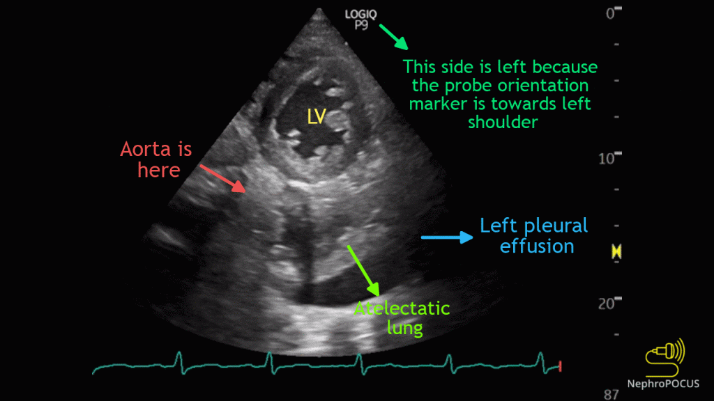

Now let us take a look at the apical window. Following images (Figures 3-5) illustrate the visualization of left pleural effusion from the apical window (actually, probe is placed slightly lateral to the cardiac apex), as well as anatomic correlation. When you see atelectatic lung, it makes the identification of pleural effusion easy. In addition, pleural effusion follows the curvature of chest wall.


In the subxiphoid cardiac view, ascites can mimic pericardial effusion. Remember you are looking at the heart through the liver in this window. When the liver is surrounded by ascites, it appears next to the heart. Below is an example showing both pericardial effusion and ascites (Figure 6). Anatomic correlation included (Figure 7). Clues to identification of ascites: intermittent appearance of the liver tissue, visualization of the falciform ligament and of course, its continuity with the peritoneum, that is visibility in other abdominal windows. Figure 8 shows another case of ascites where liver appearance is more obvious.


Right pleural effusion is frequently seen from the subxiphoid window though it typically does not mimic pericardial effusion. However, it is often confused with ascites. Figure 9 shows transverse section of the liver with an anechoic area in the posterior aspect, which corresponds to right pleural effusion. It is called ‘boomerang sign’ because of its appearance. Figure 10 is the anatomic correlate.


Figure 11 is an excellent demonstration of right pleural effusion (relatively a large one), a small pericardial effusion as well as ascites. Curvilinear transducer is used to capture a wider area.

In summary, not every anechoic thing adjacent to heart is pericardial effusion. Be aware of the anatomy and scan multiple windows to make a correct diagnosis.
Reviewed by: Sam Kant

Well, it’s a nice one, I have been looking for. Thanks for sharing such informative stuff.