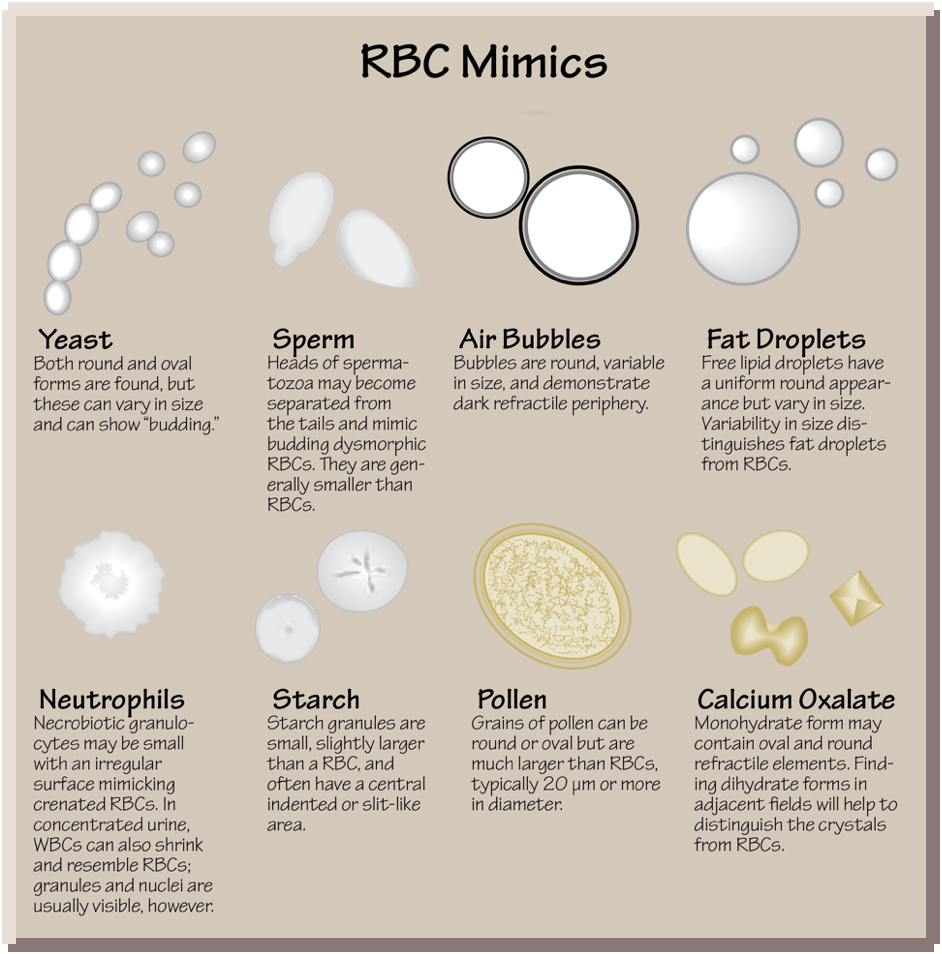
Red blood cells (RBCs) in the urine are normally 7-8 µm in diameter, circular or slightly oval and biconcave in appearance. They may appear crenated, with symmetric spiked membrane contour in hypertonic urine, or spherical, with near loss of pigmentation, in hypotonic urine. As RBCs traverse areas of damaged glomerular basement membrane, they may sustain damage which ultimately results in dysmorphic forms such as acanthocytes: ring form RBC’s with one or more cytoplasmic protrusions, or “blebs”.
Although RBCs are usually easy to identify, there are numerous RBC lookalikes which may at times be difficult to discern from RBCs. These include:
- Calcium oxalate monohydrate crystals
- Lipid droplets
- Starch granules
- Yeast
- Air bubbles
- Spermatozoa heads
- Squamous epithelial cell nuclei
- Pollen
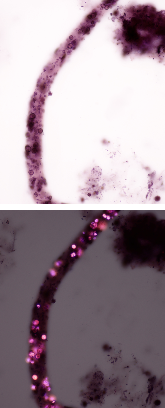
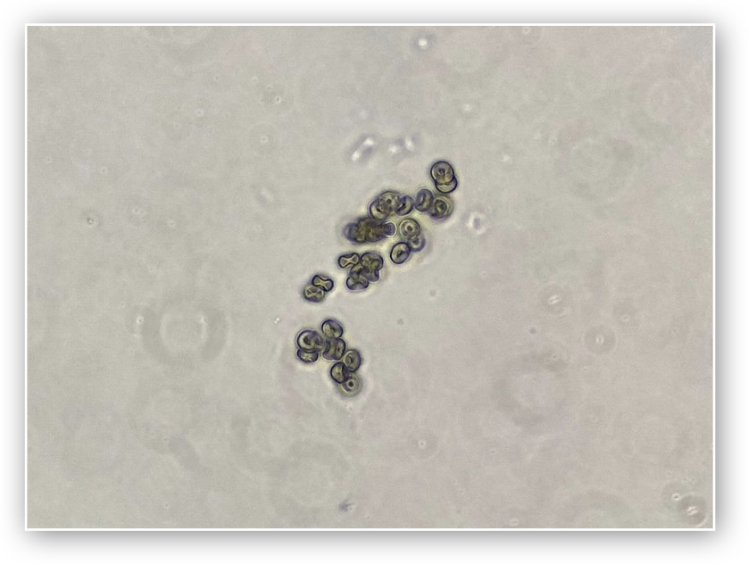
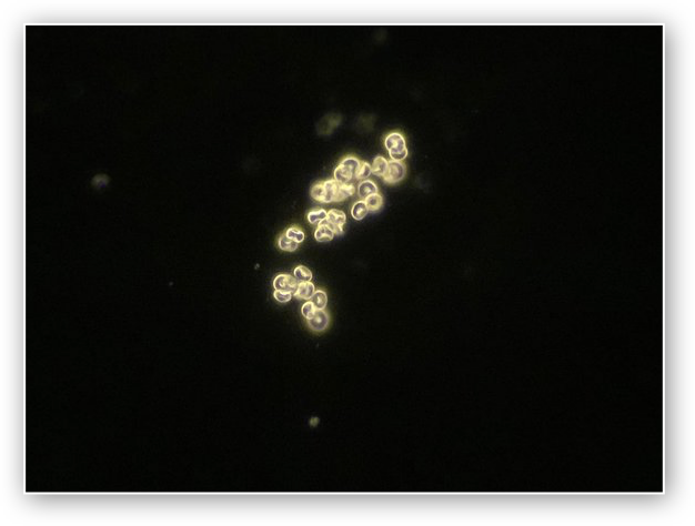
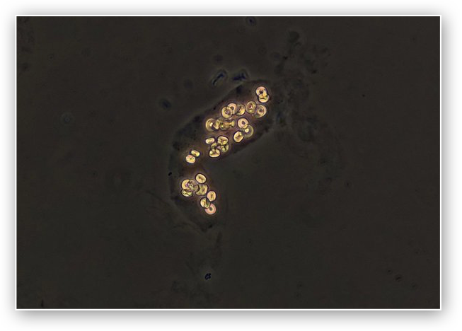
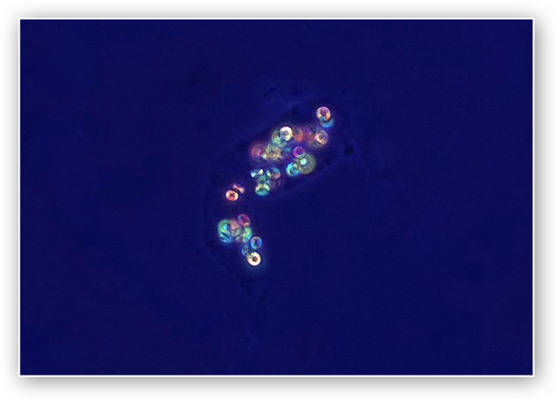
Calcium Oxalate Monohydrate Crystals
Calcium oxalate monohydrate crystal morphology is quite variable and at times these crystals may appear as small biconcave discs resembling RBC’s. When they are present within casts they are important to differentiate from RBC casts. Under polarized light they typically show polychromatic birefringence, whereas RBCs are not birefringent at all. When polarization is not available, darkfield may be useful. Calcium oxalate monohydrate crystals (as well as most crystals) have a high refractive index resulting in a “bright” appearance under dark-field illumination. RBCs under darkfield illumination appear as just a faint outline of the cell membrane.
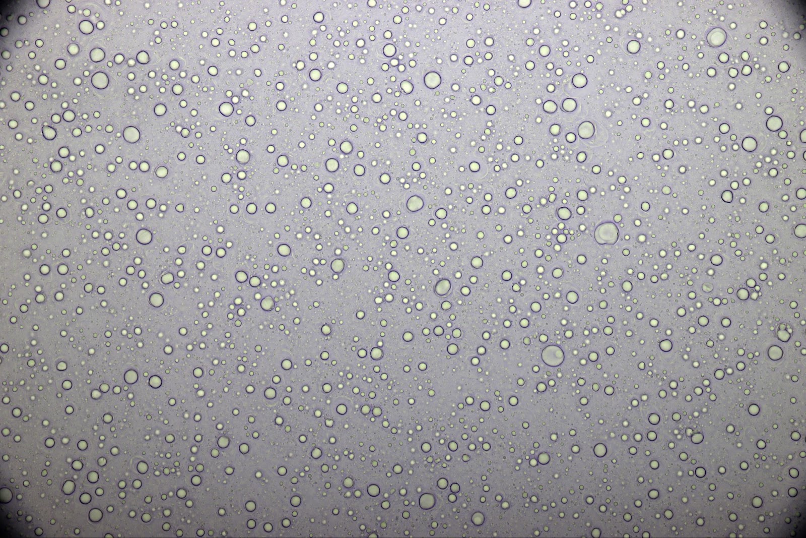
Lipid Droplets
Lipid droplets also appear as round or spherical objects but they vary in size from one droplet to another which is the main clue to their identity. In addition, under polarized light they manifest a distinct “Maltese cross” pattern of birefringence. Under darkfield illumination they appear “bright” due to their high refractive index, unlike RBCs.
Artifacts
Artifact from lotions and lubricants may appear similar to lipid droplets, spherical and with varying sizes, but they are either not birefringent under polarized light or show only a partial irregular “Maltese Cross.”

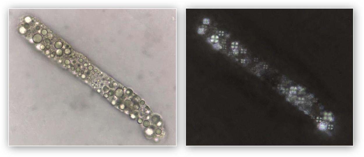
Starch Granules
Starch granules are usually slightly larger than typical RBCs and often have a central cleft. They have an irregular “Maltese cross” pattern under polarized light.
Yeast
Yeast may be difficult to differentiate from RBCs at times. Clues to yeast include a more ovoid shape as well as “budding”. Indeed “budding” yeast may mimic the appearance of acanthocytes. The opposite is true as well. In fact, urine autoanalyzers (flow- imagers and flow cytometers) may occasionally misidentify acanthocytes as budding yeast! Another clue to the presence of yeast is formation of pseudohyphae.

Other Objects Which May Appear Similar to RBCs:
- Air bubbles are round or spherical but are variable in size and have a dark distinct outline unlike RBCs.
- Spermatozoa, when the tails are no longer attached, may appear similar to RBCs but are usually somewhat smaller than typical RBCs and more ovoid.
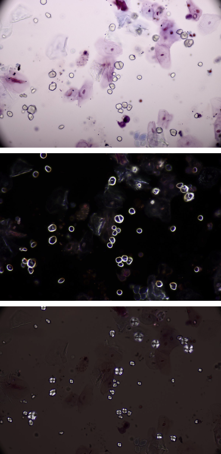
- The nuclei of squamous epithelial cells are generally the same size and shape of an RBC
- White blood cells, as they undergo degeneration may appear similar to RBCs
- Pollen grains may have a similar shape but are usually considerably larger than RBCs
Meryl H. Haber MD, David Blomerg MD, Katherine Galagan MD, Eric F. Glassy MD, Patrick C.J. Ward MB BCh – College Of American Pathologists, c2010.
In order to differentiate RBC lookalikes from red blood cells it may be necessary to use different illumination modalities (such as phase contrast, darkfield, and polarization) as demonstrated in the accompanying images.
The importance of microscopy to distinguish RBCs and RBC mimickers is important given the clinical implications that RBCs can have.
Post by: Jay Seltzer
Reviewed by: Margaret DeOliveira, Samira Farouk

I have seen a few of these in my work. Only with time, you learn to differentiate them.