“What am I looking at? ” Phagocytosis and particle internalization by cells in the urine is not common, but of clinical interest when it occurs. Phagocytosis/particle internalization can be observed in urine microscopy by the usual culprits: professional phagocytes like neutrophils and macrophages , but also by cells that we usually don’t think performs phagocytosis – that’s why the term “particle internalization” is used here.
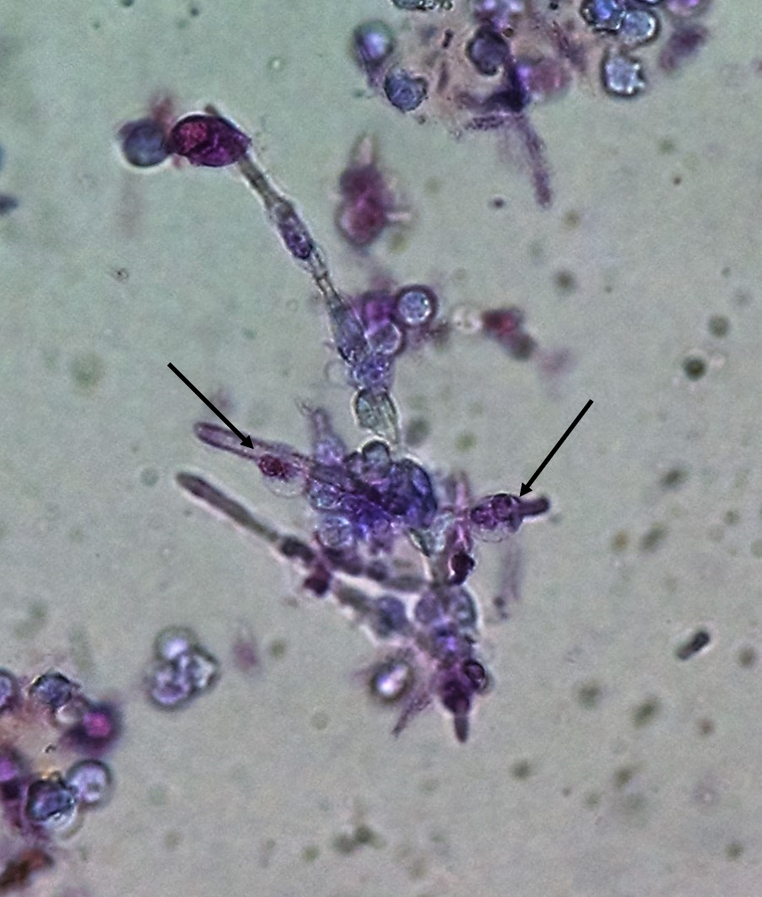
Professional phagocytes like macrophages (Fig1) and neutrophils (Fig 2) are not a surprise but are still pretty neat to see in the urine sediment and can be confusing if you don’t expect them. In Fig 2, neutrophils are taking on a fungus particle much bigger than themselves!
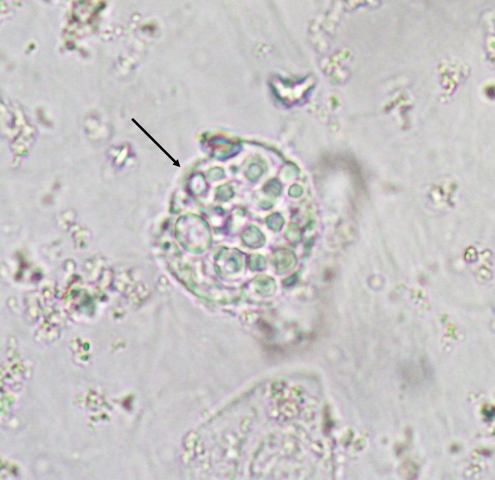
Figure 1. A macrophage with yeasts phagocytosed. The case from this particular Picture is from a patient infected with Cryptococcus neoformans, but also Candida albicans and other yeasts species can be observed. Fresh and unstained urine sediment. Bright field microscopy. Original magnification 400x. Courtesy: José A. T. Poloni.
Figure 2. Neutrophils firmly attached to a Candida sp. pseudohyphae (some pointed by arrows). It is very reasonable to assume that they are trying to perform phagocytosis of the fungal structure, despite being much smaller than fungus. Bright field microscopy. Fresh urine sediment stained with Sternheimer-Malbin stain. Original magnification 400x. Courtesy: José A. T. Poloni.
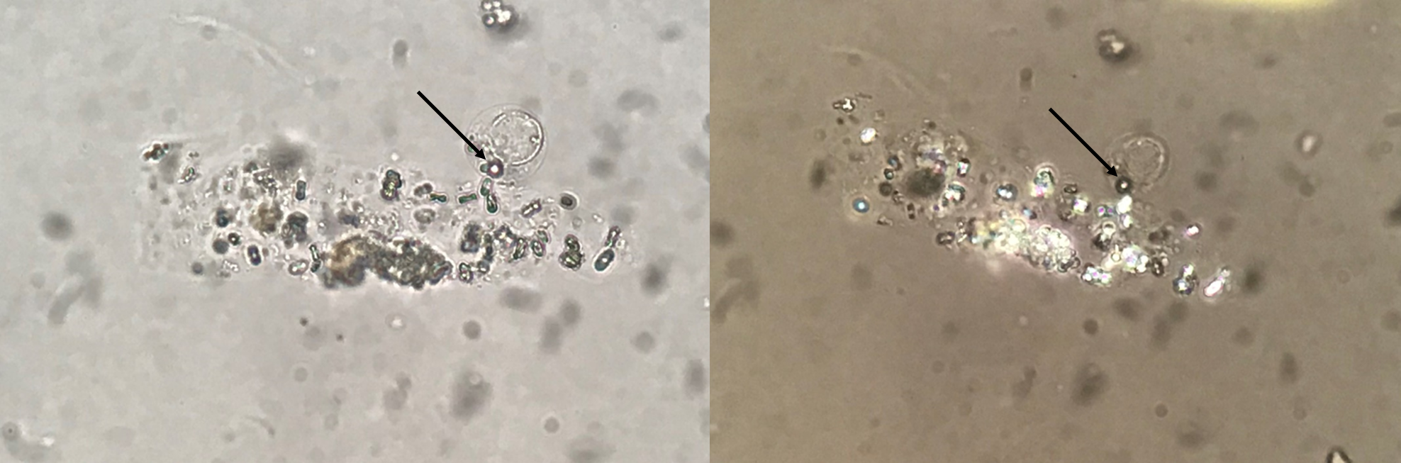
What’s less expected, however, is non-professional phagocytes like renal tubular epithelial cells (Fig. 3) doing the same job.
Figure 3. Renal tubular epithelial cell (RTEC) with calcium oxalate monohydrate (CaOxm) crystals within the cytoplasm. It is from a patient diagnosed with Primary hyperoxaluria type 1, and, on the image, the RTEC is presented with a cast containing CaOxm crystals. Fresh and unstained urine sediment. Original magnification 400x. Bright field microscopy (left) and polarized light microscopy (right). Courtesy: José A. T. Poloni
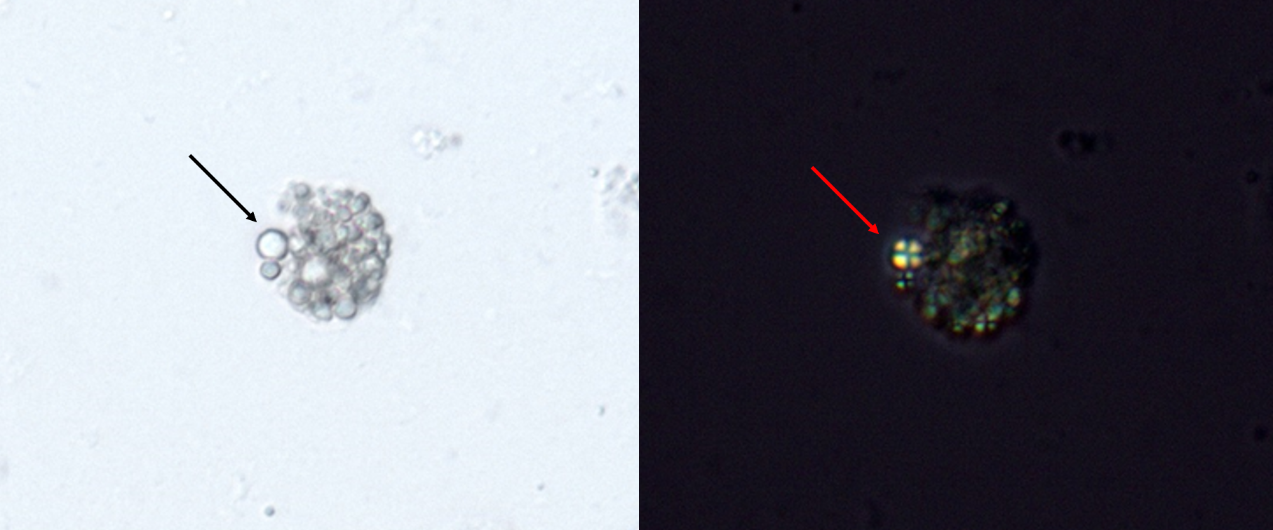
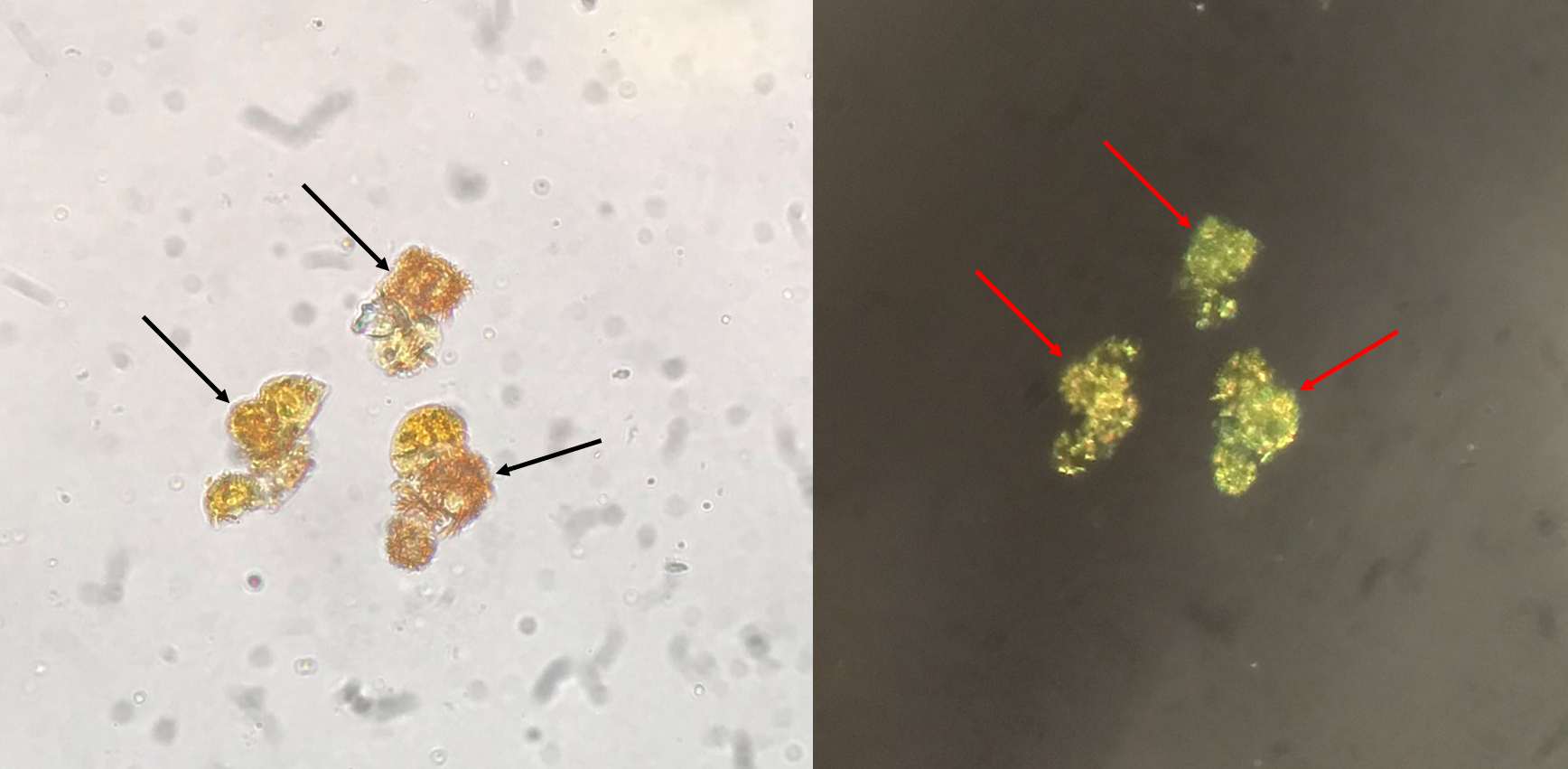
Figure 4. Oval fat body. This is probably the most known phagocytic process observed in urine microscopy. It is observed in patients with nephrotic range proteinuria. Depending on the amount of lipid droplets internalized it is impossible to identify if it is a macrophage or a renal tubular epithelial cell (thats why it is called oval fat body). Under polarized light the cholesterol esters internalized are observed forming a “Maltese cross” pattern as we see in the image on the right side. Fresh and unstained urine sediment (left) and polarized light (right). Original magnification 400x. Courtesy: Controllab
Frequently, it is not an easy task to properly identify the phagocytic/particle internalization process, because the literature has infrequent examples, usually as case reports. Also, the identification of these “particles within other particle” usually requires proper microscopic resources like polarized light microscopy (Figures 3-5) and stains like Sternhemer-Malbin stain (Figure 2) or May-Grunwald-Giemsa stain (Figure 6).
Figure 5. In patients with high levels of bilirubin, small crystals observed as needles or dots can be observed both free and within cells in urine like neutrophils, macrophages and renal tubular epithelial cells. Fresh and unstained urine sediment. Bright field microscopy (left) and polarized light microscopy (right). Original magnification 400x. Courtesy: José A. T. Poloni.
However, independently of the availability of microscopic resources or stains, we need to be able to see that there is something unusual on the slide. As we are seeing here, virtually any particle (Figures 1-8) can be phagocytosed/internalized by both professional or non-professional phagocytes, from crystals, to microrganisms, from lipid droplets to cells [5,6].
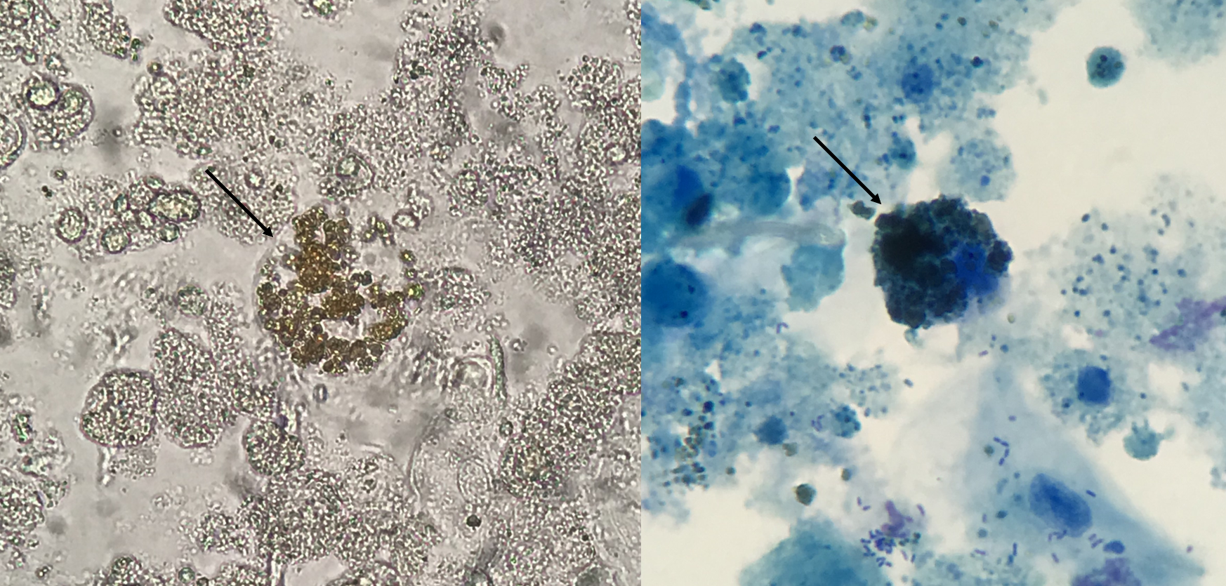
Figure 6. Hemosiderin granules observed within a renal tubular epithelial cell in the urine sediment of a patient with acute kidney injury secondary to hemolytic anemia. Fresh and unstained urine sediment (left) and fresh urine sediment stained with May-Grunwald-Giemsa stain (right). Original magnification 400x. Courtesy: José A. T. Poloni
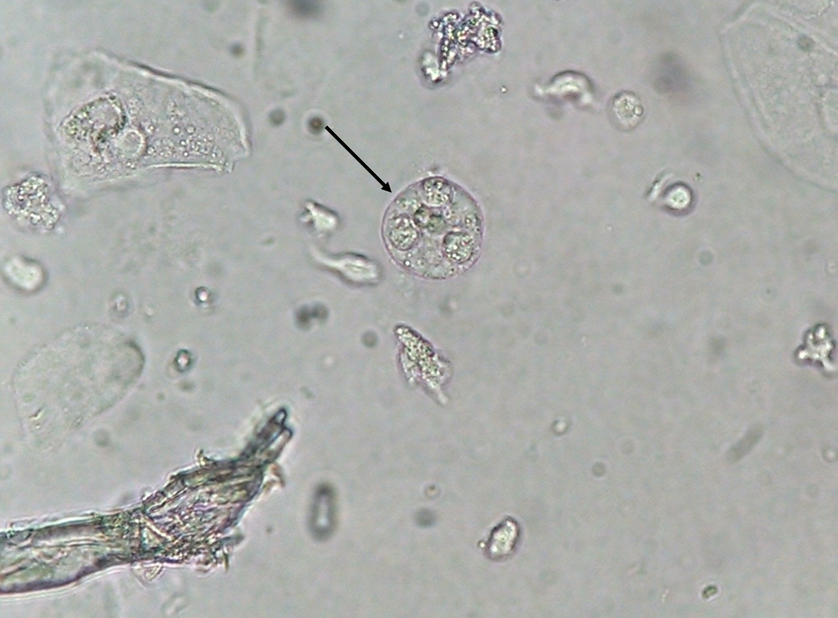
Figure 7. Neutrophils phagocytosed by a macrophage. There is no explanation known to this finding. Probably the macrophage is “cleaning” a “battlefield”, however, there is no information to prove it. Fresh and unstained urine sediment. Original magnification 400x. Courtesy: José A. T. Poloni
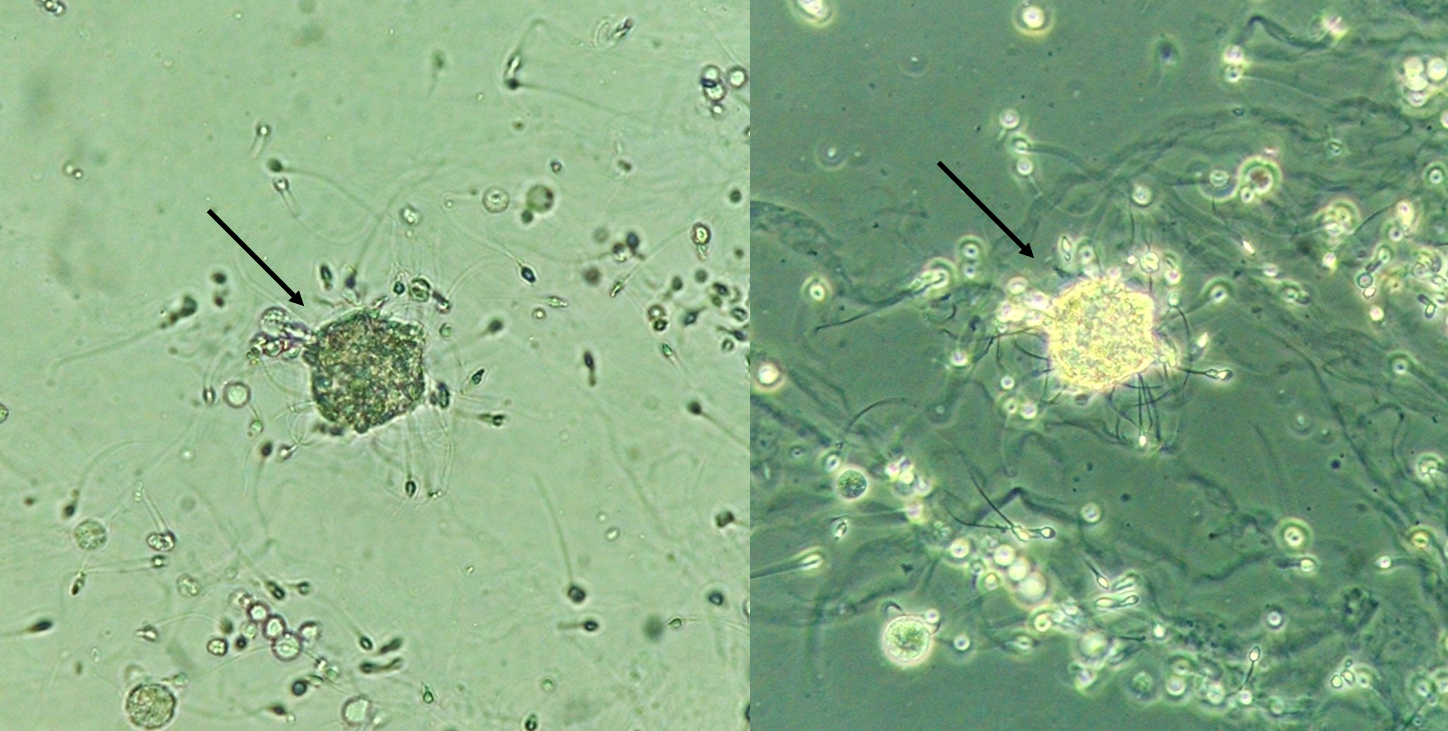
Figure 8. Sperm body. Virtually nothing is know about this urinary particle very rarely observed in the urine sediment of male patients. Fresh and unstained urine sediment. Bright field microscopy (left) and Phase contrast microscopy (right). Original magnification 400x. Courtesy: José A. T. Poloni
There is a lack of published work on phagocytosis/particle internalization in the urinalysis subject, this is surely an opportunity for professionals interested on this field to develop works and clarify these very interesting findings.
Great content! This is exactly the sort of thing I was looking for. Thanks for your help
Eduardo Ramírez
Una felicitación a todos los colaboradores de este sitio, conozco un poco del trabajo del Dr Poloni y son magníficas sus fotografías, también soy un apasionado del tema, les escribo desde México. Éxito


Great content! This is exactly the sort of thing I was looking for. Thanks for your help
Una felicitación a todos los colaboradores de este sitio, conozco un poco del trabajo del Dr Poloni y son magníficas sus fotografías, también soy un apasionado del tema, les escribo desde México. Éxito