The German Louis G.F. Thomas (1838 —1907) described “cylindroids” (Figure 1) in 1870 to indicate elongated elements partly similar to casts [1], the nature of which is still unclear at present .
There is no consensus on the definition and nature of cylindroids. According to Schreiner and Graff, cylindroids are elongated elements with a rounded end resembling a cast, and the other end resembling a mucus filament.
A prospective study of 600 sediments over a period of 4 months, Fogazzi observed cylindroids in 90 samples from 79 patients. Cylindroids were considered a morphological variant of casts by the following features:
• They are almost always associated with casts (85 out of 90 samples (94.4%).
• May have the same pleomorphism in appearance as casts (eg, hyaline, granular, cellular, fatty).
• They contain Tamm-Horsfall protein on their surface
Knowledge of the structures of the urine sediment helps in their proper identification. And since cylindroids carry the same clinical significance as casts they should not be misidentified as mucus filaments.
Phase contrast microscopy and stains (eg: Sternheimer-Malbin stain) can help with the proper visualization of cylindroids.
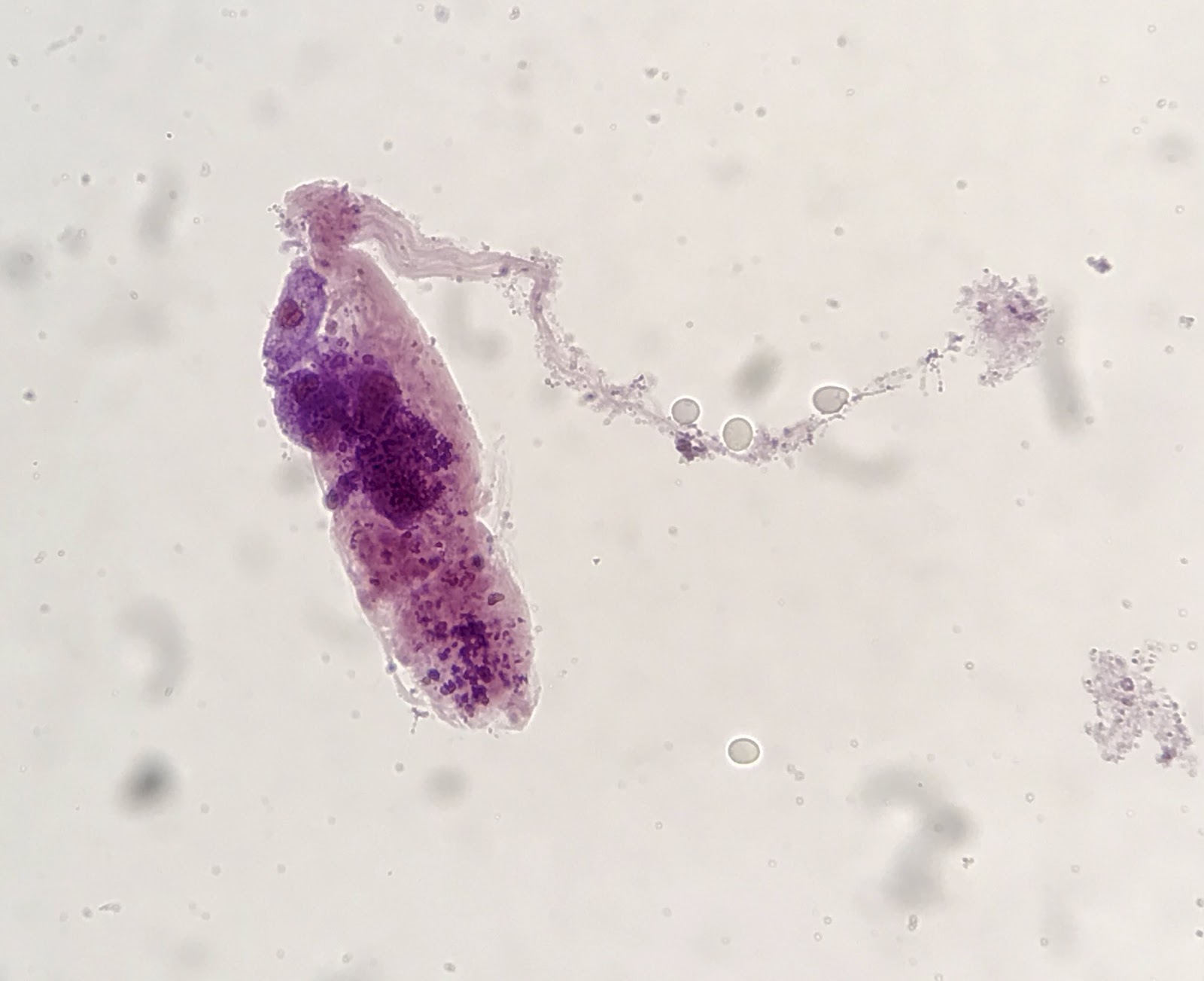
Figure 1: Cylindroid containing tubular epithelial cells and granular debris. Bright field microscopy with Kova stain (modified Sternheimer-Malbin stain). Original magnification 400x. Courtesy: Jay R. Seltzer.
References:
1-THOMAS L: Klinische Studien über die Nierenerkrankung bei Scharlach. Arch Heilkunde XI:130—156, 1870.
By: José Antonio Tesser Poloni
Reviewed by: Margaret DeOliveira, Jay Seltzer, Samira Farouk