Waxy casts are found during microscopic examination of the urinary sediment. Like other casts, they are cylindrical structures, but they are distinctly characterized by a high refractive index. Their shape can be straight (Fig. 1A, top cast), convoluted (Fig. 1B), or curvilinear. Their surface is typically smooth with “melted wax” appearance (Fig. 1A, bottom cast) which can be better appreciated at low powerfield (Fig. 1C & Fig. 1D). However, they can also be filled with granular material (Fig. 1A, top cast) or contain embedded cells. The edges can be intact or cracked and are easily visible by adjusting the fine focus knob on the microscope.
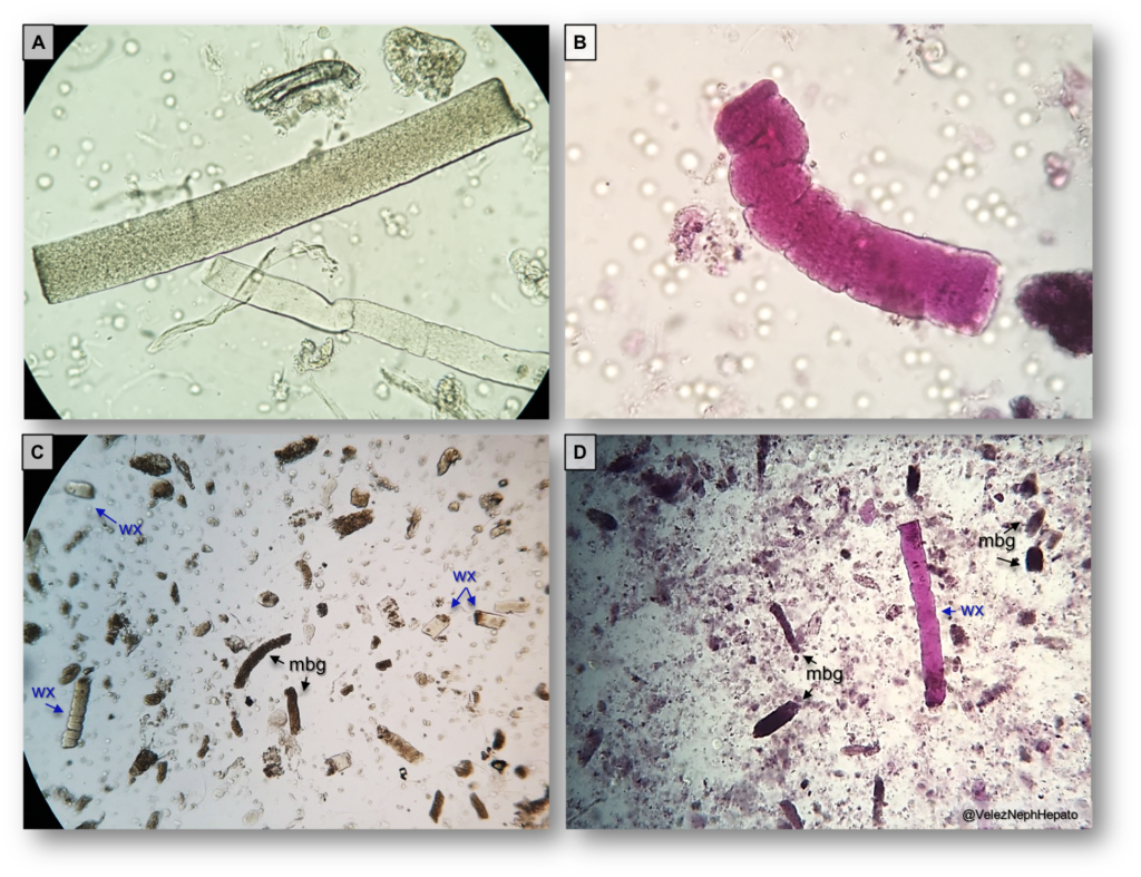
“Broad” waxy casts are those with greater width that are thought to form at the convergence of collecting ducts near the papilla. Despite their early description, the composition of waxy casts remains unknown. It is postulated that they originate from granular casts. While most textbooks highlight them as being indicative of chronic kidney disease, waxy casts are commonly seen during severe acute tubular injury, intermixed with muddy brown granular casts (Fig. 1C & Fig. 1D).
All images were captured with bright field microscopy. Specimens shown in panels B and D were treated with Sternheimer-Malbin stain.
Post by: Juan Carlos Velez, MD
Nephrologist, @OchsnerNephro