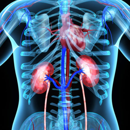
If you are like me, you vaguely know about renal scintigraphy but not so much about the details of the radioactive agents or the utility of the scan itself. Basically, renal scintigraphy is a 2D nuclear medicine scan of the kidney. “Scintigraphy” is a a technique which refers to the use of a detector with a radioactive tracer to obtain an image of a bodily organ or a record of its functioning. Images appear similar to a pulmonary ventilation/perfusion (V/Q) scan, and this form of kidney imaging can be used for a multitude of indications: most notably, estimating glomerular filtration rate (GFR), visualization of renal cortex, assessment of the impact of hydronephrosis, collecting system leaks, and more as described below.
Before we start, let’s meet the three teams of radioactive agents that are used: Filtered, Secreted, and Retained
1. FILTERED agents are used to estimate glomerular filtration rate.U
- 125I-Iothalamate (IO) – weak photon energy source; not very useful
- 51Cr-Ethylenediaminetetraacetic Acid (EDTA) – closest to inulin clearance; not available in the US
- 99mTc-diethylenetriaminepentaacetic Acid (DTPA)
- Correlates well with EDTA for GFR > 30 mL/min
2. SECRETED agents are used to assess renal perfusion by estimating effective renal plasma flow and obstruction. The term “effective renal plasma flow” is preferred as the dose must be delivered to the kidney and extracted.
- 99mTc-Mercaptoacetyltriglycine (MAG3) –used to assess “functional obstruction” and also the preferred agent for captopril renography in patients with impaired renal function because looks at parenchymal retention, but also used in normal renal function
- Can confound images as partially seen in hepatobiliary system and gut
- 99mTc-L,L- and D,D-Ethylenedicysteine (EC) – possibly superior to MAG3
- 99mTc-(CO3)Tricarbonylnitriloacetic Acid (NTA) – still under investigation, possible less hepatobiliary or gut activity
3. RETAINED agents are retained through proximal tubular receptor mediated endocytosis and are helpful for static cortex imaging.
- 99mTc-Dimercaptosuccinic Acid (DMSA) – uptake is dependent upon renal blood flow, GFR, and proximal tubule endocytosis
- 99mTc-Glucoheptonate (GH) – cannot use for diuretic renography as filtered, then retained
Let’s move on to the the 4 different types of renal scintigraphy studies:
- Dynamic Renal Scans
Dynamic scans can be used to evaluate kidney function and perfusion. There are three ways to assess GFR: plasma-based, by measuring change in plasma levels of the radioactivity; camera-based, where a computer takes sequential images as the radioactive agent clears the body and uses a software to assess GFR; and quantitative indexes (relative uptake of a radiopharmaceutical over time, radiopharmaceutical time to peak, or ratio of excretion of a radiopharmaceutical).
Filtered or secreted radiopharmaceuticals may be used for dynamic renal scans. These scans may also provide a split renal function, also called differential renal function (relative percentage of each kidney’s contribution to the GFR). These studies may also reveal urinary leaks via extravasation of the radioactive agent.
2. Static Kidney Cortex Imaging
Renal cortex imaging appears to be more important in the pediatrics world and is used to identify ectopic kidneys, renal agenesis, horseshoe kidneys, and multicystic dysplastic kidneys. It is also used in pediatrics for the diagnosis of pyelonephritis, as it is more sensitive than ultrasound.
“Cold defects” are radiopenic areas (regions that do not “light up”) in the cortex and may represent scar, pyelonephritis, cysts, tumors, infarct, or remnants of prior trauma. These scans can also identify split function as well.
3. The Loop Diuretic Renogram
Not all hydronephrosis means loss of function, and this test may aid in understanding of the functional impact of observed obstruction. In pediatrics, pelvic ureteral junction anomalies usually resolve, and only 25-30% result in parenchymal damage, so identifying the right patient to intervene upon is critical. In the adult world, there are patients with known longstanding hydronephrosis and slowly declining renal function in the setting of multiple comorbidities, it is hard to know what to do with the hydronephrosis.
After the loop diuretic (typically furosemide) is given, the radioactive agent is traced. Retained or slow movement of the tracer suggests loss of function. The furosemide dose is usually 1.0 mg/kg, with a maximum dose of 40 mg in adults (20 mg in children). Higher doses are needed in obesity, chronic diuretic use, hypoalbuminemia, or impaired kidney function as organic acids competitively inhibit transport of furosemide. Interestingly, MAG3, the radiopharmaceutical used, shares the same transporter as furosemide in the kidney as it is protein bound. 10-15% of studies may be difficult to interpret because of intermediate diuretic response.
4. The Captopril Renogram
Although controversial, this scan is used to identify patients who may benefit from renal artery stenting, monitor stented patients, or identify ischemic nephropathy or partial renal vein thrombosis.
The captopril renogram may be used a data point to help decide if stenting may be helpful, keeping in mind that up to 25% of normotensive patients over 50 years old will have evidence of atherosclerotic renal artery stenosis and indiscriminate stenting is not recommended. Ultrasound can achieve similar findings as scintigraphy, but is also operator dependent and may be unreliable in obese patients. The gold standard for diagnosis is angiogram.
In appropriately screened individuals with preserved renal function, the sensitivity and specificity of this test was over 90% each. However, almost 50% of studies will give an indeterminate probability result, especially in patients with renal dysfunction. Results come back as low (less than 10%), intermediate, or high (greater than 90%) probability of renovascular hypertension.
The test is conducted by administering an angiotensin converting enzyme inhibitor (usually captopril 25-50 mg) and then 1 hour later injecting a radiopharmaceutical agent (MAG3 or DTPA). The symmetry of uptake of the tracer, time to peak uptake, and retention are then analyzed. It is imperative the technician monitors blood pressure as asymptomatic hypotension can lead to abnormal results.
Finally, are there limitations to renal scintigraphy?
Of course. There are many limitations which can result in false positives or equivocal results. Patient movement can degrade already poor resolution images and enhance background noise. Choice of radioactive agent is usually performed by the radiologist and is important to ensure results are informative (ie DMSA cannot be used to assess GFR because it is retained in the kidneys). Recent urinary tract infections may reduce uptake of DMSA, and food intake 4 hours prior to a captopril renogram may delay gastric absorption and confound results. Additionally lasix renograms can be influenced by poor response to furosemide or poor elimination of radioactive agents because of back-pressure from distended native bladders, reflux from neobladders, or dilated urinary systems resulting in a reservoir effect. Reflux from neobladders are more commonly seen in ileal conduit systems. Medications such as angiotensin converting enzyme inhibitors (ACEi), angiotensin II receptor blockers (ARBs), and non-steroidal anti-inflammatory drugs (NSAIDs) affect effective renal blood flow and confound results.
So, what is it good for?
In the era of modern imaging, renal scintigraphy is still a useful tool in the provider’s armamentarium. In my opinion, it is probably most useful is to exclude disease states such as renovascular hypertension and obstruction, urinary leaks, or assess differential kidney function. Keep in mind that any test is limited by technician and radiologist familiarity and expertise. In general, the more precise you are in your clinical question, the more helpful the study will be.
Post by: Amy Yau, MD
Nephrology fellow, Icahn School of Medicine at Mount Sinai


Excellent summary 👍