White blood cells (WBC’s) in the urine may occur as a result of infection or inflammation anywhere in the urinary tract and the segmented polymorphonuclear neutrophil is the type most often encountered. These WBC’s are identified by the presence of granular cytoplasm and a segmented multi-lobed nucleus. Sternheimer-Malbin (SM) staining facilitates visualization of the segmented nucleus but specific identification may require focusing up and down as the segments may exist in different focal planes. WBC size averages 10-12 µm in diameter and, while these cells are usually spherical, if they remain in contact with glass for a period of time they may form pseudopods and appear elongated or amoeboid.
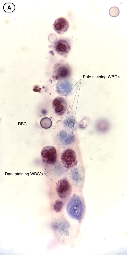
There are two different WBC staining patterns observed with use of the SM stain. Dark staining WBC’s are characterized by translucent or granular cytoplasm and avid magenta or red staining of the segmented nucleus. Pale staining WBC’s have pale indistinct blue staining of the cytoplasm and nucleus and may have visible cytoplasmic granule movement.

WBC staining pattern depends on age and viability of the cell (pale staining WBC’s are young and remain viable while dark staining WBC’s represent older non-viable cells). The presence of cytoplasmic granular motility in viable WBC’s depends on relatively low viscosity of the cytoplasm, as occurs in the presence of hypotonic urine.
Pale staining WBC’s exhibiting granular motility may be called “glitter cells”, Sternheimer-Malbin cells”, or “granular motility cells”. Although they were once thought to be pathognomonic for pyelonephritis they are a non specific finding.
C WBC cast containing both pale and dark staining WBC’s (brightfield with SM stain) ![]()
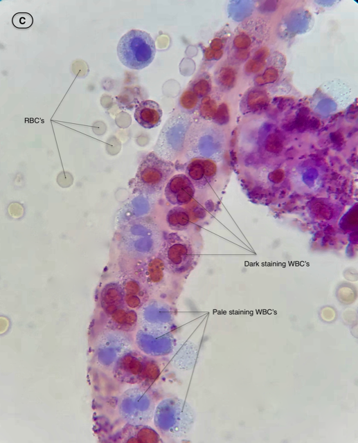
D. Pseudocast (brightfield with SM stain). Note what looks like a cast is actually just WBC’s aggregating along mucus threads

WBC’s may enter the tubules as a result of inflammation in the glomerulus or interstitium. Formation of WBC casts are thus indicative of an inflammatory process such as glomerulonephritis or interstitial nephritis. It is worth pointing out that WBC casts, while often thought of as pathognomonic for interstitial nephritis, are a common finding in the urine sediment of patients with proliferative glomerulonephritis.
Tip: Beware of pseudocasts. WBC’s often occur in clumps and may be elongated or aggregate along mucus threads giving the appearance of a cast. Note that in these cases there is no tubular protein matrix encasing the cells.
Post by: Jay Seltzer, MD