BK virus (BKV) is a DNA virus that infects up to 90% of the adult population. After initial exposure in childhood, the virus becomes latent in urothelial tissues. In transplant patients, BKV can reactivate in the context of potent immune suppression which may result in uncontrolled viral replication and eventually BKV-associated nephropathy (BKVAN). Prompt detection of viral reactivation can improve outcomes and therefore active screening after transplantation is recommended.
Screening tests for BKV infection include:
–identification of decoy cells (DCs)
-qualitative and quantitative polymerase chain reaction (PCR) test to detect BKV DNA in urine and plasma
-urine electron microscopy
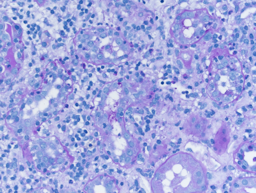
-allograft biopsy (gold standard method).
Once diagnosed with BKVAN, between 16% and 50% of transplant recipients will lose the graft.
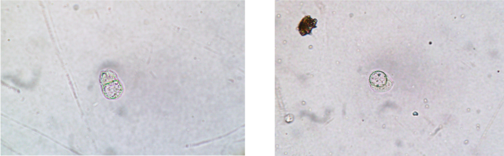
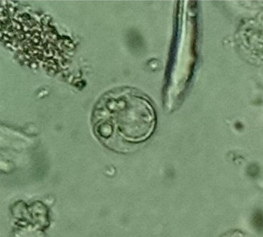
Decoy cells are renal tubular epithelial cells and other uroepithelial cells that manifest changes associated with viral infection. They appear in the urine presenting as glassy appearing, homogenous intranuclear inclusions.
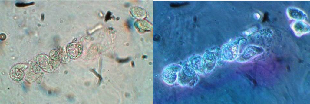
The classical description of the morphology of the DCs, consists in nuclear enlargement, which confers a ground glass appearance and displacement of the nucleus toward the periphery of the cell as if the nucleus were escaping from the cell (comet-like morphology); chromatin margination, which is chromatin clumping along the nuclear membrane; abnormal chromatin patterns; single nuclear inclusion body surrounded by a peripheral halo, which confer a bird’s eye appearance to the cells; and cytoplasmic vesicles (Figures 1 – 3). These cellular changes are suggestive of BKV infection. However, decoy cells are not entirely sensitive or specific for BKV infection. Renal tubular epithelial cells (RTEC) casts with features of BKV infection (Figure 4) were observed on urine microscopy in seven of eight patients with biopsy‐confirmed BKVAN.
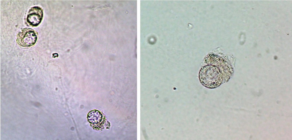
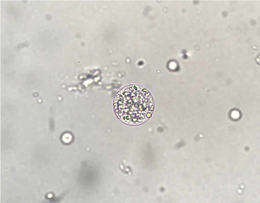
An important point to mention is the fact that the urine sediment profile of patients presenting DCs features commonly RTECs (Figure 5) and macrophages (Figure 6) that can be mistaken as as DCs.
Decoy cells can be easily detected during routine urinalysis performed by motivated professionals. It can lead to an early detection of BKV reactivation in the population of kidney allograft recipients helping on the management of these patients preventing than to develop BKVAN.
Post by: José Antonio Tesser Poloni
References:
Fogazzi, G. The Urinary Sediment (2010, Kidney International)
Reilly Jr. RF and Perazella MA. Nephrology in 30 Days (2014, Lange)