Hyaline casts are traditionally known to lack clinical significance. However, because they are frequently found during urinary sediment microscopy, hyaline casts should be correctly identified and distinguished from other types of casts. They are colorless transparent cylindrical structures with round edges that are essentially composed solely of a matrix of Tamm-Horsfall (uromodulin) mucoprotein. At times, a few cells or debris can be seen “trapped” within the cast.
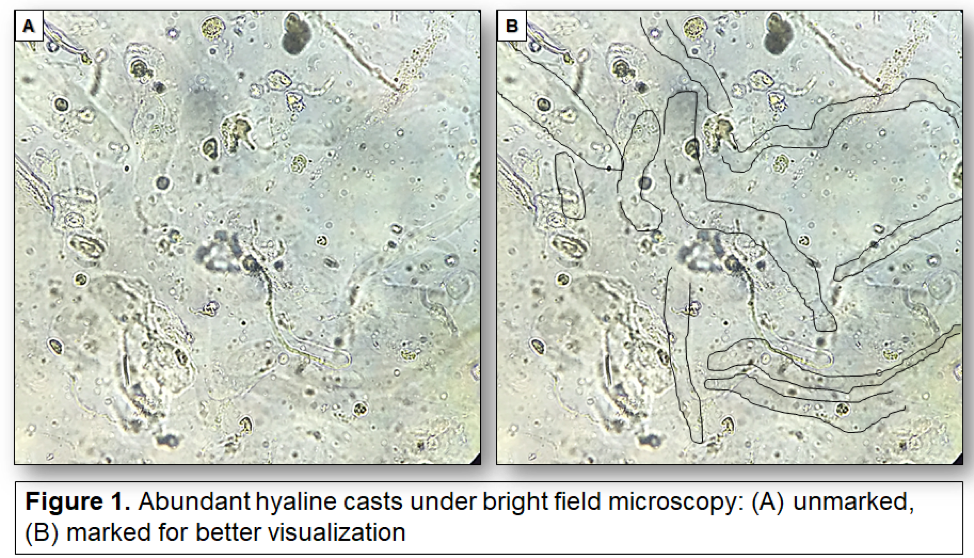
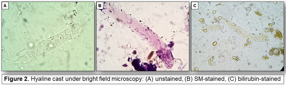
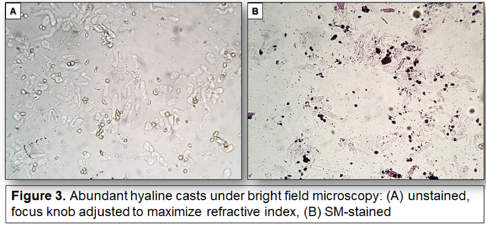
Because of their low refractive index, they can be easily missed when inspected under bright field microscopy (Figure 1). The use of Sternheimer-Malbin (SM) stain allows for better visualization (Figures 2,3). They are also better seen in hyperbilirubinuria (Figure 2). Adjusting the focusing knob can also help visualizing them (Figure 3). Phase contrast microscopy is best suited for easier recognition of hyaline casts (Figure 4).
Hyaline casts have been reported in variety of physiological and pathological settings, such as exercise, dehydration, following intravenous diuretic administration, etc. In the context of disease, hyaline casts can be seen alone or intermixed with other types of casts and/or cells.
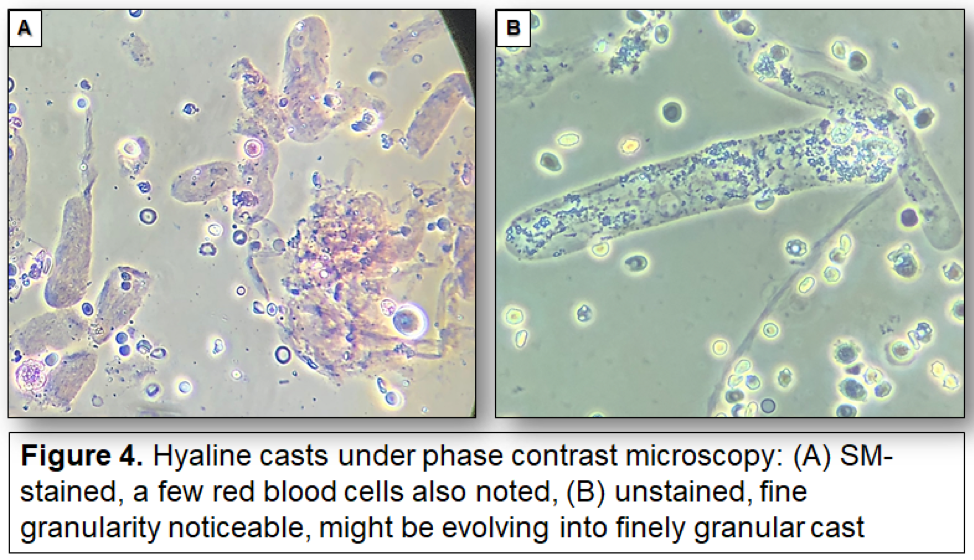
Overall, they are thought to denote presence of a sluggish tubular flow regardless of the cause (functional or intrinsic) and can be present both in acute and chronic kidney disease, although they do not point to any specific etiology or disease process. Occasional granularity might be observed (Figure 4), but when the granules are prominent, the cast should be called finely granular instead.
Juan Carlos Q. Velez, MD
#PisseProphet #UrinarySediment #UrineMicroscopy #SpinYourOwnUrine
@OchsnerNephro
