Last week we covered 5 clinical scenarios involving arteriovenous fistula and graft complications. We now discuss another 5 common scenarios as part 2 of the series.
Please note: these cases are vignettes created for educational purposes and patient consent has been obtained by author for clinical images.
Case 1
70 year old ESKD patient on dialysis for past 10 years. She has required multiple fistulograms and angioplasties on her right upper extremity fistula, including transposition. She presents with fevers, chills and drainage at the access site. Wound and blood culture are growing MRSA. Her condition deteriorates in the hospital and she requires transfer to ICU for septic shock. Her AVF is shown:
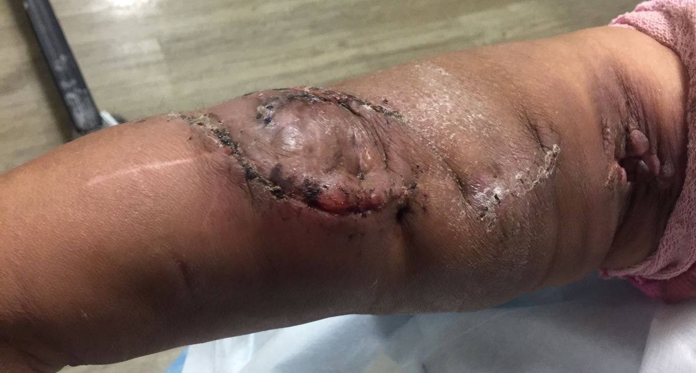
What do you observe?
Swollen, erythematous access with crusted drainage. It is tender on palpation.
What is the plan of action?
Emergent surgical removal of the AVF , I&D around the fistula site and establishment of alternative dialysis access
Diagnosis: Infected AVF
Case 2:
A 40 year old female on home hemodialysis for past 5 years was referred to your vascular lab for fistulogram since she is having prolonged bleeding and poor clearance on dialysis. You notice the following on her access:
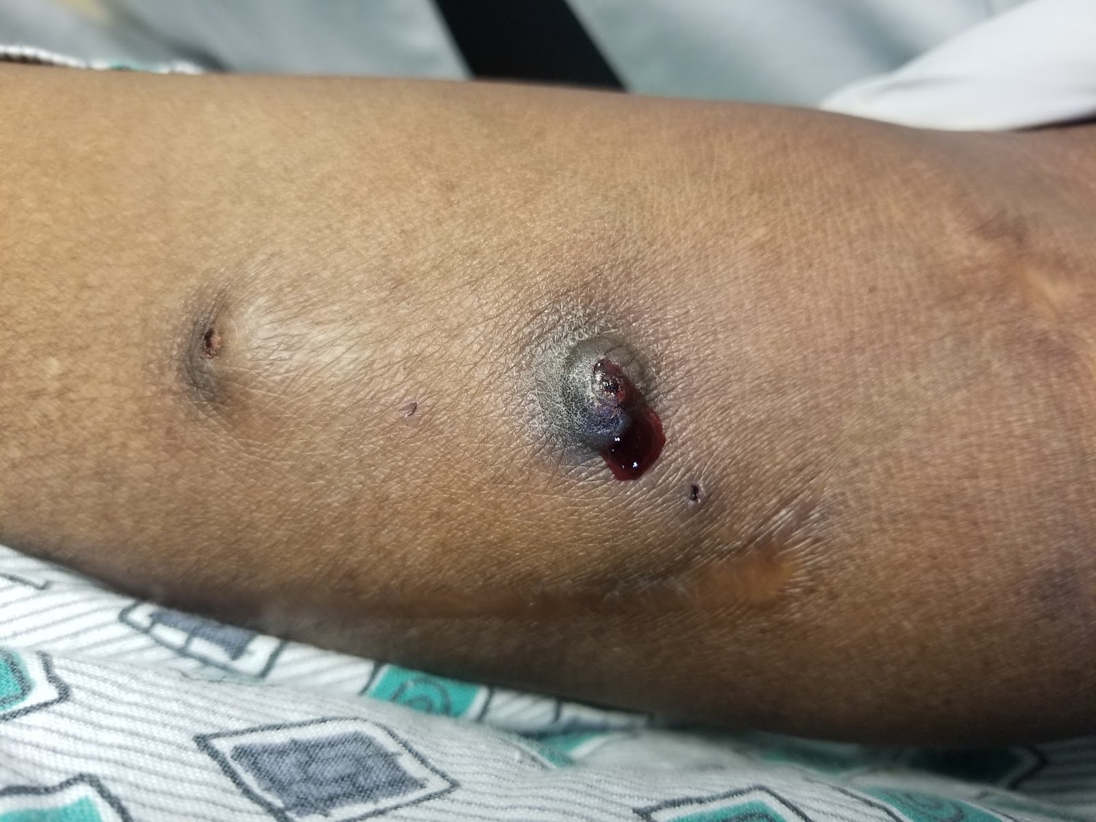
What is her cannulation technique?
The patient is using the buttonhole technique. Instead of sharp needles, dull needles are placed in the exact same spot at every cannulation, creating a ‘tunneled track’. Over time cannulation is less painful, however risks of infection are higher especially due to staph species.
What is wrong in this picture?
The scab on her button hole came off and she is bleeding. There is a swelling at the buttonhole site.
What should be done next?
Fistulogram should be cancelled and she should be sent to the ER. If the button hole track opens up completely she may exsanguinate. An ultrasound study needs to be done to rule out abscess or hematoma formation at the site.
Diagnosis: Buttonhole infection/hematoma formation
Case 3:
50 year old male on in center hemodialysis through right upper extremity AV fistula for the past 12 years. This is his 4th admission this year for ‘volume overload’, hypoxic respiratory failure and decompensated heart failure. His cardiomyopathy is non-ischemic. He has no known intrinsic pulmonary process. Rolling the patient’s sleeve reveals this AVF:
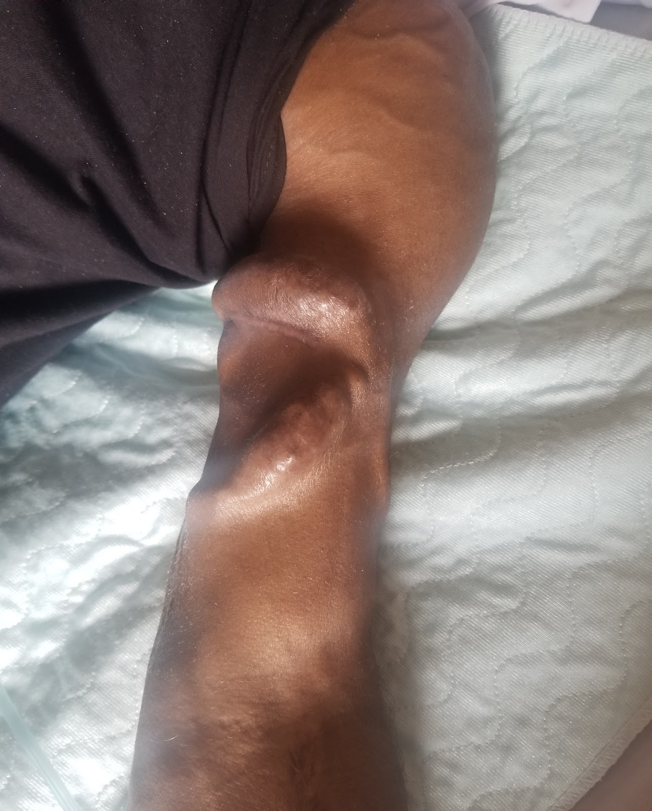
What does this appearance raise suspicion for?
A dilated, tortuous AVF with very brisk flow should raise concern for high output cardiac failure related to the AVF, especially if other causes of volume overload are ruled out (cardiac, pulmonary, liver)
What maneuver would you elicit on physical exam?
Nicoladoni Brenham Sign: On occlusion of the AVF, there is a rise in systolic bp by ~20 mmHg and a drop in pulse rate of ~20 beats /min
What diagnostic test should you order?
AVF flow studies and Echocardiogram. If flow through fistula > 30% cardiac output it is hemodynamically significant. Occlude fistula and check cardiac output during Echo – if cardiac output drops significantly, there is indication for banding/ligation of fistula to decrease flow through the access.
Diagnosis:
High output heart failure related to AV – fistula (You can read more about this condition on our NSMC blog post Size and Flow Matter – Memoir of an AV fistula)
Case 4:
You are assessing a dialysis patient in the ER. Her fistula is as shown below. She has had the fistula for 10 years and has not had any issues with blood flows on dialysis. She achieves adequate KT/V and Urea reduction ratio. Over the years the fistula has developed areas of dilatation as shown below. Her last dialysis was 2 days ago and she ran a full 3.5 hour treatment without any difficulty.
What should you do next?
Perform a physical exam and assess for outflow stenosis. Aneurysmal dilatation of an AVF should raise suspicion for obstruction to blood flow proximally.
What diagnostic test should you order?
Although not emergent, this patient will need a fistulogram to assess for central vein stenosis once outflow tract obstruction is ruled out on physical examination.
What precautions should be taken?
Dialysis nurses should be advised to avoid cannulating the aneurysmal areas. The size of the aneurysm should be measured and documented for future references.
What are the warning signs?
Rapid increase in size, shiny and pulsating overlying skin, pain, infections.
Diagnosis:
Stable Aneurysm of AV fistula.

Arteriovenous Grafts
The next section deals with complications associated with AV grafts (AVG). AVGs are used for patients who do not have adequate native veins for creation of an AVF. AVGs are usually the second best choice of vascular access creation. As with AVF’s, AVGs can be created in the upper limbs and the thighs (last resort).
Complications associated with AVGs are almost similar to the ones with AVFs:
1.Venous anastomotic site stenosis due to neointimal hyperplasia.
2.Development of Pseudoaneurysms.
3.Thrombosis.
4.Central vein stenosis.
5.Infections
Recognition of those complications are similar to our previous cases. Let’s however highlight a very common complication that many patients with AVG present with.
Case 5
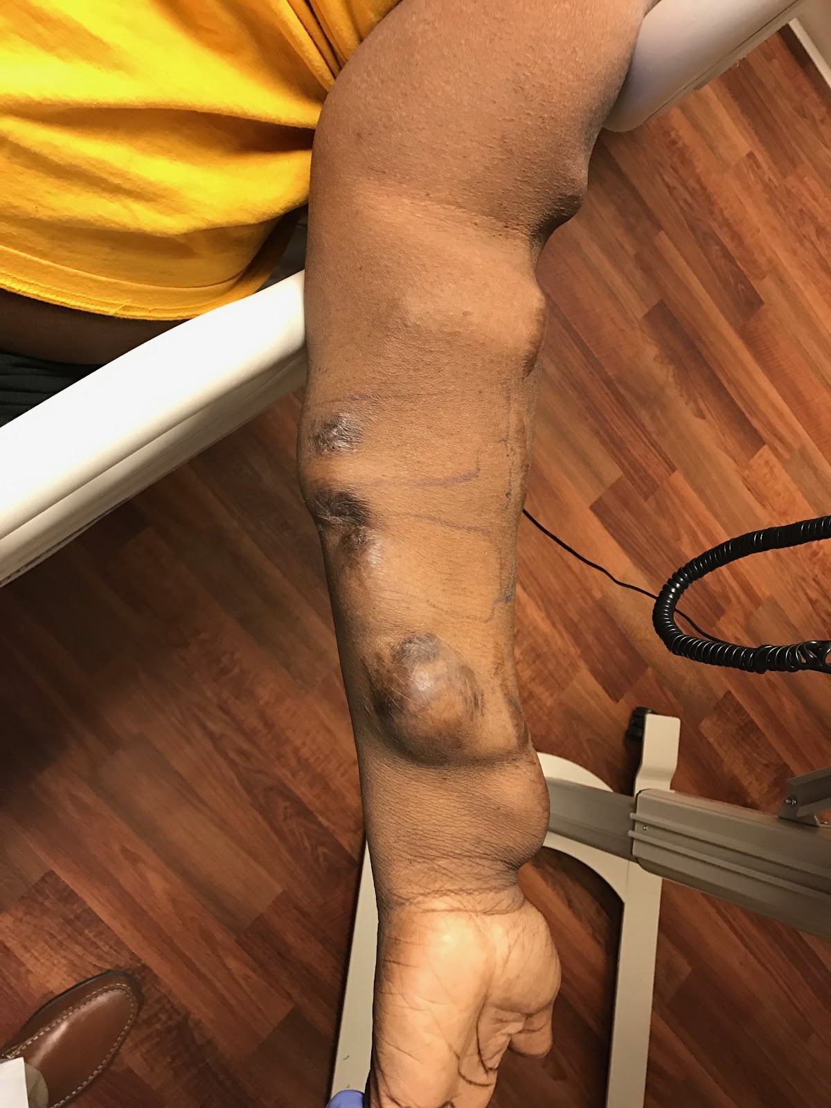
A 50 year old male is being assessed in the hospital prior to dialysis. He has a left forearm AVG that was placed about 6 years ago. He reports no issues on dialysis, but does mention he has had ‘swelling’ on his graft for the past couple of years.
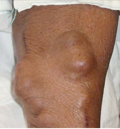
What can you observe?
Pseudoaneurysmal dilatation of the AVG at cannulation sites.
Why is it called ‘pseudo’?
AVGs develop pseudoaneurysms at frequently punctured sites. The high blood flow in the AVG causes extravasation of blood in a cavity outside the graft but confined next to it by surrounding tissues.
Why is this a problem?
Pseudoaneurysms can cause thrombus formation due to turbulent flow. There is also higher risk of bleeding during cannulation at those sites. Monitoring is similar to AVF aneurysms. Surgical intervention is required in larger pseudoaneurysms or rapidly enlarging ones
Diagnosis:
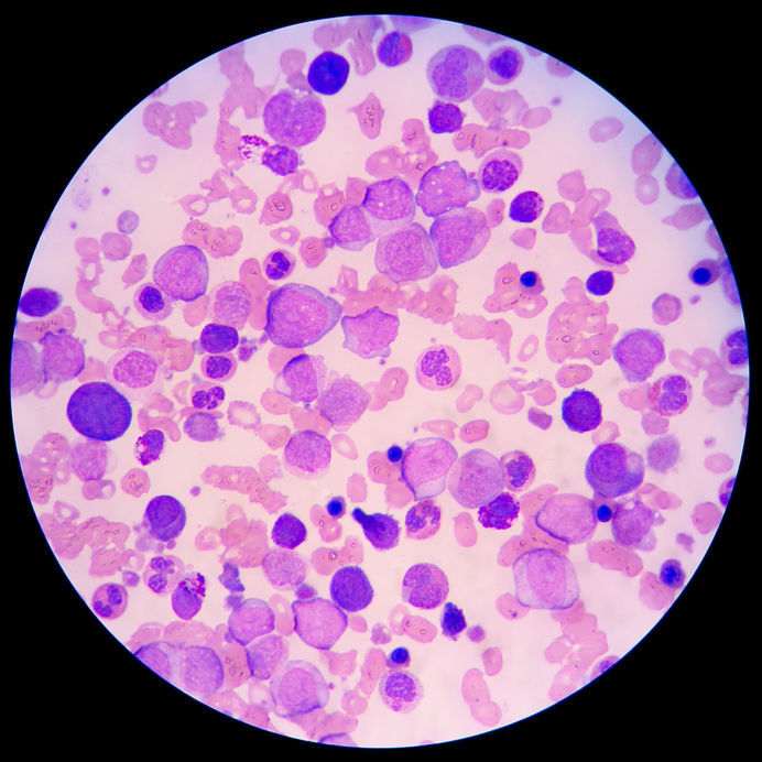
Pseudoaneurysm related to AVG. Appearance of pseudoaneurysm during ‘shuntogram’ is shown below (white arrows)

Post by: Bhavnish Bucktowarsing, MD
ASDIN fellow
Acknowledgments: This post is part of a collaboration between the Renal Fellow Network and the American Society of Diagnostic and Interventional Nephrology (ASDIN), whose mission is to provide excellence in dialysis access care to improve outcomes for patients with kidney disease. Special thanks to Tushar Vachharajani, Aisha Shaikh, Edgar Lerma, and the Education Committee of ASDIN for their comments and suggestions for this post. For more information about the ASDIN mission or membership, click here. We would also like to thank Anil Agarwal, Robin Shah, Nabil Haddad, Khaled Boobes for assisting with this post.

These excellent case studies simplify learning what to look for when assessing the av access.
Thanks for sharing
very helpful & simplified