 Friday we did two biopsies on the Consult Service…one of which turned out to be a fairly interesting diagnosis of fibrillary glomerulonephritis.
The case in brief: a 71 yo woman with a history of HTN and no diabetes with a history of CKD and a Cr which has steadily been going up over the past 6 months with a Cr which is now 1.9. Her urine sediment has been persistently positive for red and white blood cells. A serologic workup has been negative including a negative SPEP and UPEP.
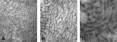
The biopsy showed variably involved glomeruli with a mesangioproliferative pattern of injury and some mild to moderate interstitial fibrosis. IF staining was remarkable for patchy IgG staining and approximately equal kappa and lambda staining. The electron microscopy findings–essential for the diagnosis of fibrillary GN–showed the accumulation of 20nm non-branching, randomly arranged fibrils which are ultrastructurally similar to amyloid fibrils but differ by virtue of their larger size and lack of reactivity to Congo Red staining.
Many patients with fibrillary GN progress to ESRD. Unfortunately there are no therapies available other than standard CKD management with ACE-I/ARB and BP control. Often the disease is idiopathic though there has been an association with hepatitis C infection.

