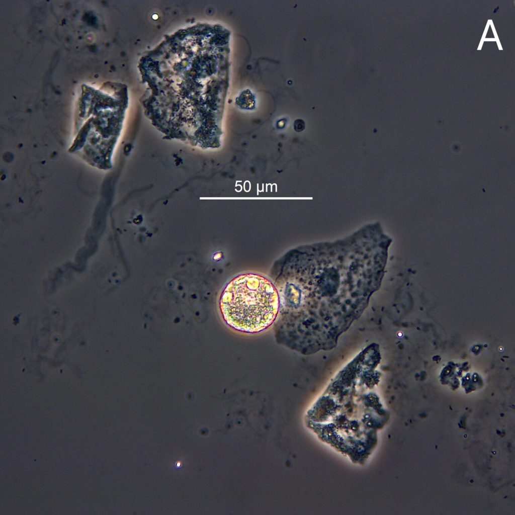
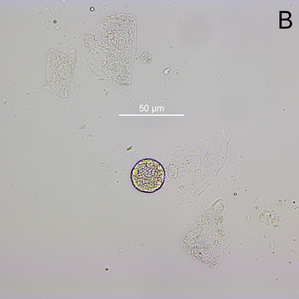
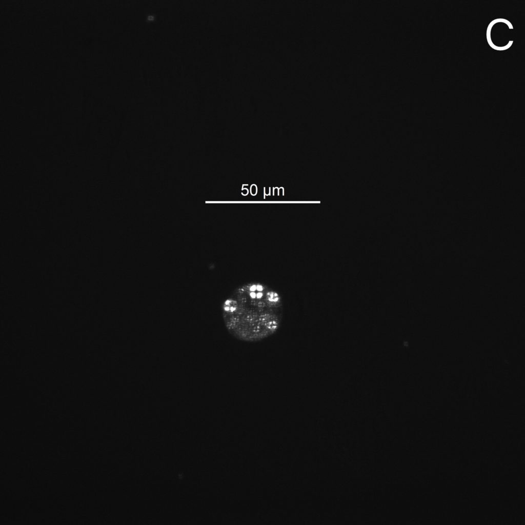
Below, take a look at nearly round “oval fat bodies” in the urine of a patient with the nephrotic syndrome. Their presence is a sign of higher grade proteinuria:
The so called “oval fat bodies” are a common finding in the sediment of patients with higher grade proteinuria. They represent desquamated tubular epithelial cells or macrophages that are full to the brim with lipid droplets. The cellular lipid accumulation probably results from uptake of free fatty acids (FFA) complexed to albumin. These FFA are readily metabolized to triacylglycerol and cholesterol esters and stored in lipid droplets, possibly to protect the cells from harmful effects of free fatty acids.
Given their pretty unique morphology, identification of oval fat bodies by phase contrast (Fig. A) or bright field (Fig. B) microscopy is usually straightforward. Presence be confirmed by demonstrating the typical birefringence (“Maltese cross”) of lipid droplets under polarized light (Fig. C). The histologic equivalent to oval fat bodies are tubular epithelial cells with fatty changes or foam cells of monocyte/macrophage lineage.
Post by: Dr. Florian Buchkremer (@swissnephro)


very interesting, i love looking at this urine sample os much. thank u for what u do this is very beneficial to me and my fmily rn