Several types of organisms can be identified in the urine sediment. Bacteria, fungi, and parasites are 3 classes of microorganims that can be found.
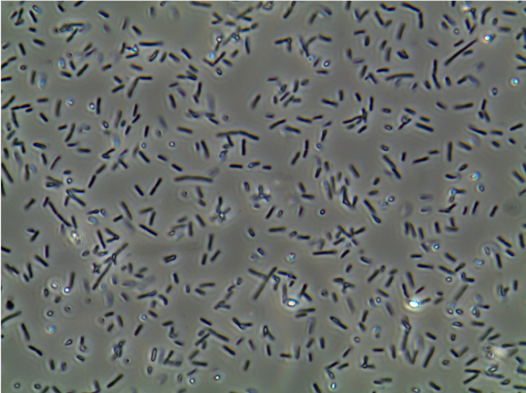
1. Bacteria
Rods and cocci can be identified in the urine sediment and urinary tract infection (UTI) can be suspected if bacteria is observed in samples recently obtained by the clean catch technique, particularly if associated with pyuria. However, bactiuria and pyuria may be the result of a contaminated sample. In a patient with vaginal discharge and vaginitis squamous epithelial cells may be seen with or without fungi or Trichomonas vaginalis.
2. Fungi (Yeasts)
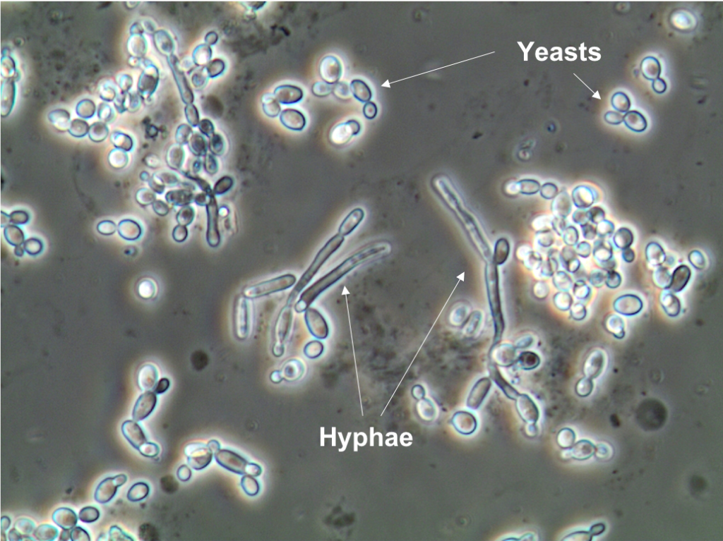
Fungi (yeasts) are unicellular organisms that reproduce as budding yeasts with the separation of the sister cells. Candida is the yeast type more commonly observed in urine, with Candida albicans most commonly identified . There are more than 80 Candida species, but only a few are pathogenic to humans. Of note, red blood cells (RBC) and yeasts have similar size and morphology. The most common cause of Candida in urine sediment is sample contamination with vaginal discharge in a patient with vaginitis. Candida may also lead to fungal UTIs – specially in patients with diabetes mellitus, those with structural abnormalities of he urinary tract, and in patients on antibiotics or immunosuppressants. Candida can be associated to invasive candidiasis that can lead to uretritis, cystitis or pyelonephritis infection (casts containing yeast may be found in the urine)
3. Parasites
2 species of parasites have been described to be found in the urine sediment: Trichomonas vaginalis and Schistosoma haematobium.
Trichomonas vaginalis (Figure 3) is a flagellated protozoa that is slightly larger than a polymorphonuclear white blood cell (WBC). The main characteristic of this protozoa is the presence of five flagella; one of them folded back and attached to the parasite body by an undulating membrane. When alive, they are easy to identify due to their active, irregular and fasts movements throughout the slide. When dead, however, they are very hard to distinguish from polymorphonuclear WBCs. When observed in the urine sediment they are usually linked to sample contamination by genital secretions. Trichomonas vaginalis is commonly present in the urine sediment with squamous epithelial cells, polymorphonuclear WBCs, bacteria, or Candida.

Below is a video showing Trichomonas in a urine sample (original magnification 400x):
Trichomonas vaginalis (culture sample). Bright field microscopy. #parasitology pic.twitter.com/9NZfwxEyWV— José A. T. Poloni (@JoseTesser) September 12, 2019
Urinary schistosomiasis is endemic in several geographic regions (Middle East, particularly in the Nile River valley, West and South of Africa, and also some areas of the Arabic peninsula) affecting approximately 100 million people. The infection with Schistosoma haematobium is acquired when a person is in contact with a source of water contaminated with cercariae (larval stage), that are able to penetrate the skin and migrate throughout the bladder venous plexus where mature females releases eggs. The presence of eggs stimulates ureteric granuloma formation comprised of macrophages and eosinophils, leading to mucosal hyperemia and ulceration. Hematuria that is the main clinical manifestation of Schistosoma haematobium infection and can lead to subsequent obstructive uropathy. Observation of the parasite’s eggs in urine may be the clue to detect the infectious process.
References:
1. Recomendações da Sociedade Brasileira de Patologia Clínica/Medicina Laboratorial (SBPC/ML): Realização de exames em urina. Editora Manole. Primeira edição. Barueri-SP, 2017.
2. Fogazzi GB. The urinary sediment an integrated view. Elsevier. 3rd edition. Milan, 2010.
Post by: Jose Antonio Poloni Tesser