Ultrasound features of Kidney Cysts

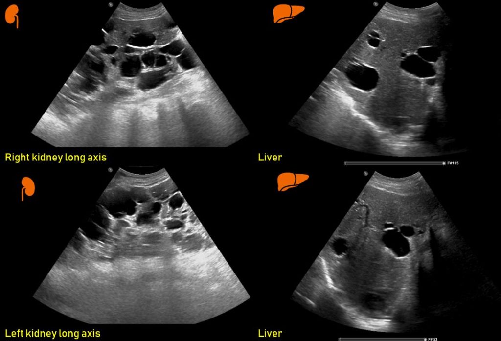
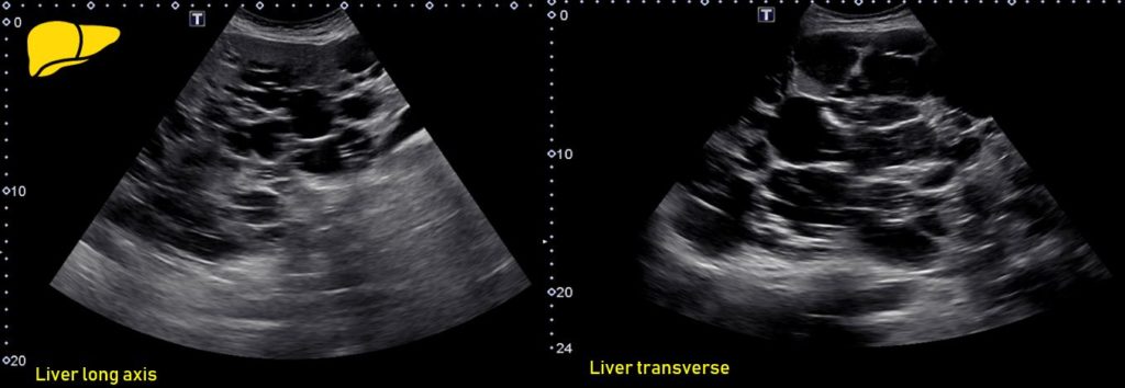
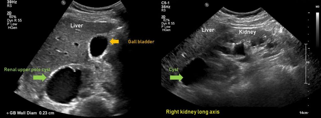
Kidney cysts are commonly encountered in clinical practice- overall prevalence has been estimated to be 10.7%. Here are some common sonographic scenarios:


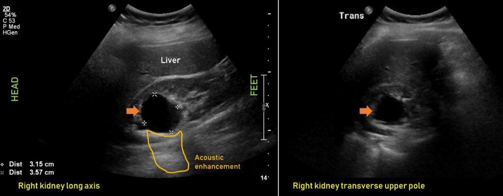
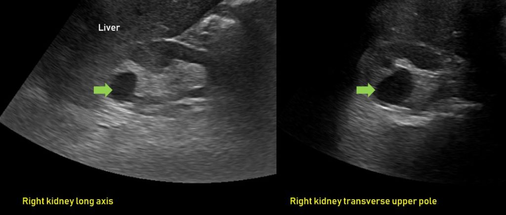
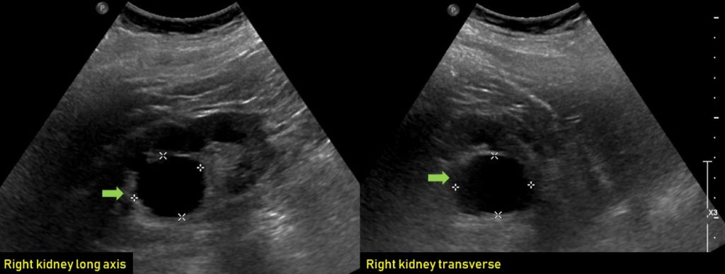
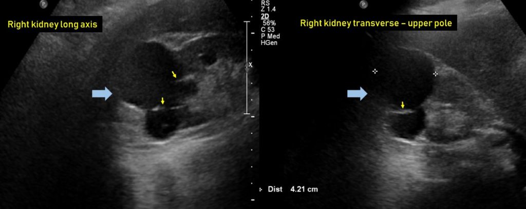
Figure 1: A simple cyst in the upper pole of the right kidney. It is anechoic without any internal echogenicities. Note the acoustic enhancement, which is an artefact that helps identify cystic structures.
Figure 2: A simple cyst in the upper pole of the right kidney.
Figure 3: An exophytic simple cyst in the lower pole of the right kidney.
Figure 4: A small cyst (c) in the right kidney with possible calcification (arrow). Asterisks (*) indicate ‘edge shadowing artefact’, which means a narrow, hypoechoic, shadow line extending distal to the lateral edges of the fluid-filled cavity. It is a refraction artefact.
Figure 5: A small subcentimeter simple cyst in the right kidney. This is very obvious because the surrounding kidney is bright (increased gain/brightness of the image) but can be missed if proper sweeping motion is not performed to visualize the whole kidney while acquiring the image.
Figure 6: A large simple cyst in the middle of the right kidney. This is called parapelvic cyst and can be confused with hydronephrosis.
Figure 7: A large simple cyst in the left kidney. Note that in the upper left image, the cyst is not visible. If proper sweeping movements are not performed, these kind of posterior cysts can be missed despite being big. The location can be better appreciated on the CT scan (lower right panel).
Figure 8: A huge cyst that appears like a urinary bladder at first glance. Note that it is connected to upper pole of the left kidney.
Figure 9: A small but complex cyst with internal echoes in the right kidney. Note the thin parenchyma as well as increased cortical echogenicity compared to that of liver suggestive of chronic kidney disease. Asterisks indicate small amount of ascites.
Figure 10: A large cyst in the left upper pole with possible calcifications (bright spots). Note that calcifications may or may not give acoustic shadowing depending on their size.
Figure 11: A complex cyst with septations in the right upper pole.
Figure 12: A huge hemorrhagic (layering echogenic debris) cyst in the lower pole of the left kidney. Note that ultrasound cannot definitively diagnose hemorrhage. A CT scan would be helpful in this regard.
Figure 13: Renal milk of calcium cyst: refers to the viscous colloidal suspension of calcium salts found either within a calyceal diverticulum or within a simple renal cyst. This calcium typically moves with patient’s position.
Figure 14: Polycystic kidneys and liver. Presence of liver cysts in addition to renal cysts is a clue to the presence of autosomal dominant polycystic kidney disease.
Figure 15: Extensive liver cysts in a patient with autosomal dominant polycystic kidney disease.
Figure 16: Right upper pole cyst can be confused with gall bladder in off-axis views. Knowledge of the anatomy and obtaining multiple images in different scan planes is the key.

Abhilash Koratala, MD

Leave a Reply