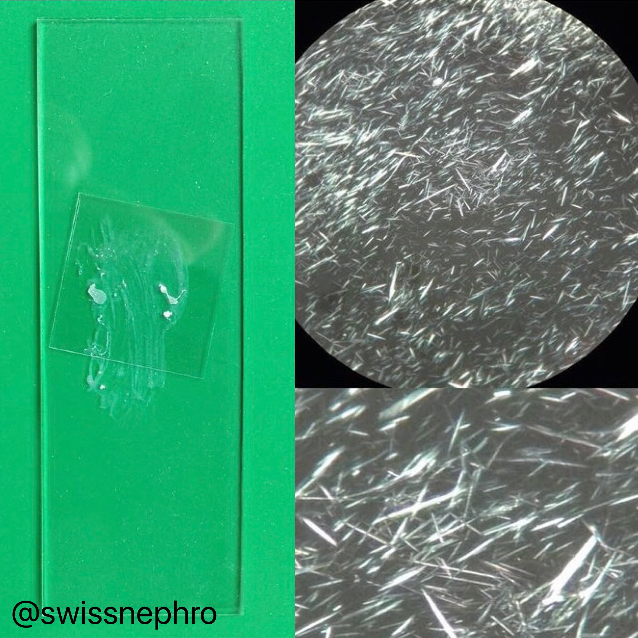
One of the welcome side-effects of doing your own urine sediment examination is having easy access to a microscope and some basic lab equipment. This allows you to examine all sorts of things besides urine (Fig. 1).
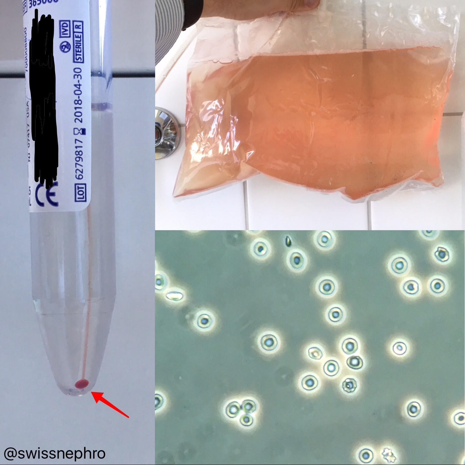
For me, this is especially useful for peritoneal dialysis fluid samples. I like to look at them myself because I get almost immediate results and a better feel for what is going on within the peritoneum.
WBCs (Fig. 2), bacteria (Fig. 3) and RBCs (Fig. 4) are all readily identified without any special staining, just as in urine.
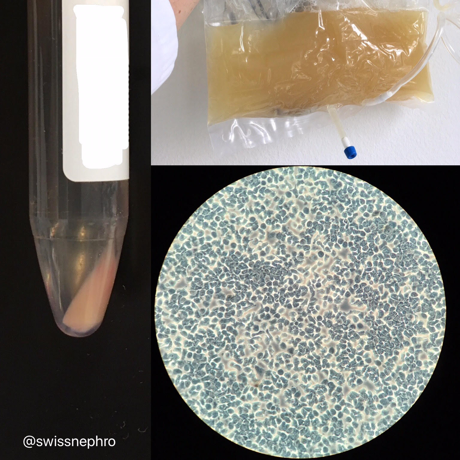
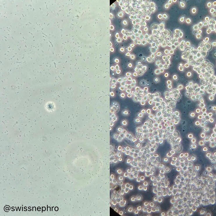
Figure 3. Peritoneal fluid sediment examination in a patient with PD-related peritonitis. Left, ~1h after symptom onset, showing abundant cocci in chains and only one (mesothelial?) cell. Right, ~6h later, now with scores of leucocytes. (Phase contrast, original magnification X 400)
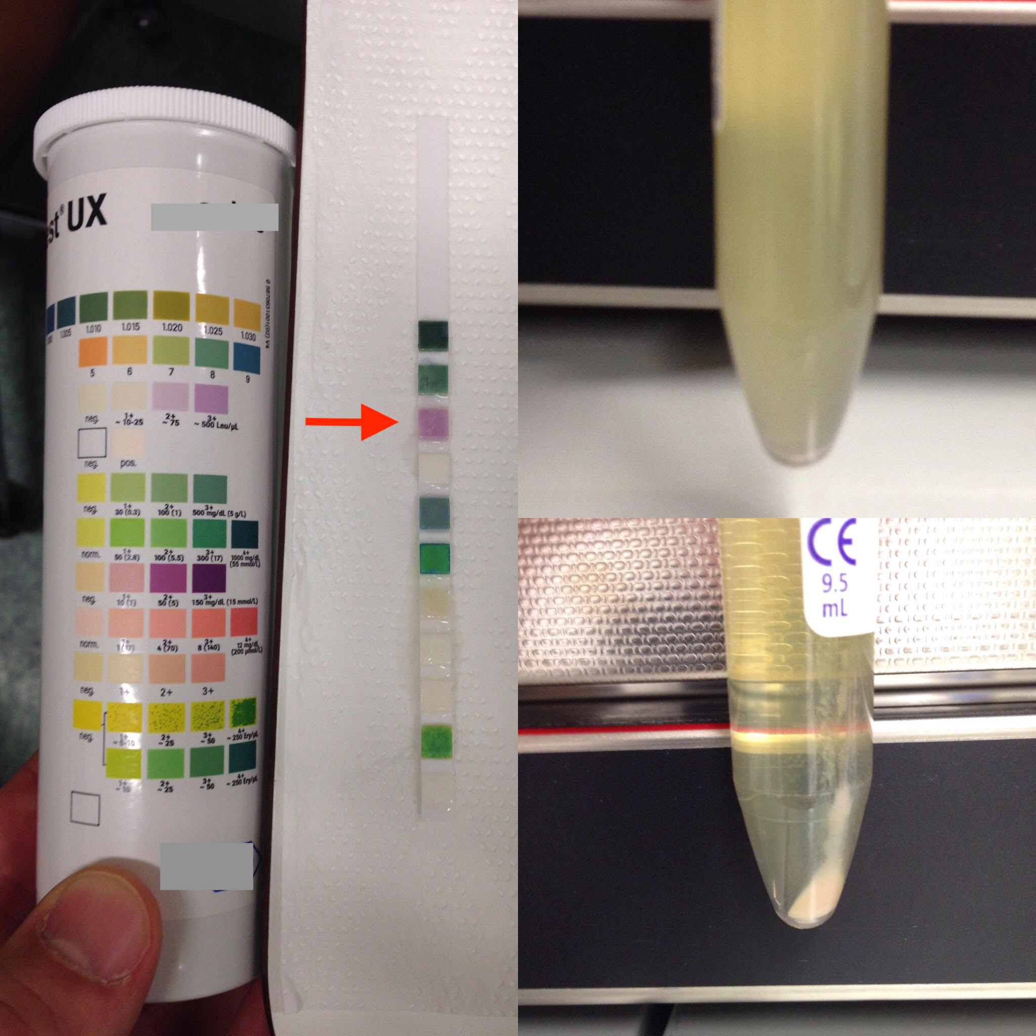
The urine dipstick works on this fluid as well (Fig. 5).
Of course, this basic examination is only a bedside supplement to formal laboratory evaluation (e.g. WBC differentiation, exact quantification, Gram staining).
Post by Florian Buchkremer
Edited by Anna Gaddy

