We’re all taught that looking for dysmorphic red blood cells on urinalysis is a useful marker of glomerular hematuria. But how do we define “dysmorphic”? And how was this association originally studied? One landmark paper was this 1991 study in Kidney International by Kohler et al.
The authors initially make the point that the term “dysmorphic red blood cells” encompasses a wide range of RBC morphologies that may be seen in the urine. This includes discocytes, echinocytes, etc etc (shown below)–all of which are made possible by the deformable membrane of the RBC, necessary for its ability to navigate through very narrow capillaries.

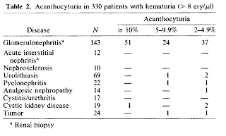
In the paper, the authors look at the urines of 351 patients with hematuria, 143 of which had biopsy-proven glomerulonephritis and the rest of which had hematuria from other diseases (e.g., AIN, cystic kidney disease, nephrolithiasis, etc.), as well as controls from non-hematuric healthy individuals.
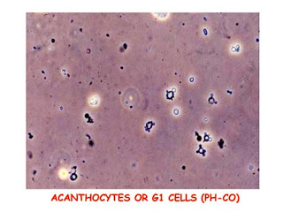
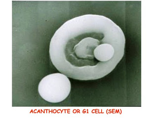
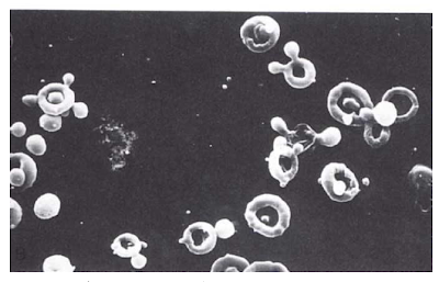
Acanthocytes (ring-shaped RBCs with blebs of membrane coming off–sometimes described as RBCs with “Mickey Mouse ears”) were the best predictor of glomerular disease compared to all other dysmorphic RBC types. Overall, acanthocytes appeared in 12.4% of all excreted RBCs in cases of biopsy-proven hematuria, and were very rarely seen in non-glomerular disease and controls. At least 5% acanthocyturia was noted in 75 out of 143 GN patients (giving a sensitivity of 52%) and in 4 out of 187 patients with nonglomerular disease (giving a specificity of 98%). The sensitivity of acanthocyturia for detecting glomerular disease could be increased by examining more than one urine sample.
Other types of dysmorphic cells (e.g., echinocytes, etc) were present in glomerulonephritis at greater levels than acanthocytes, but were also commonly found in non-glomerular kidney disease and thus are not specific. Furthermore, the number of echinocytes, discocytes, and stomatocytes were found to change when the pH, osmolarity, or protein content within a urine specimen was varied, whereas the number of acanthocytes remained relatively constant when these variables were altered.
Here are some really nice pictures of acanthocytes, employing either phase-contrast microscopy or scanning electron microscopy.


correct me if i have a mistake I understood that isomorphic erythrocytes are red blood cells with a normal shape and with the condition of ph and urinary osmolarity change their shapes either phantom or crenulated and are considered non-glomerular bleeding while dysmorphic red blood cells are of glomerular origin and acanthocytes are included in this group of erythrocytes
At any rate, I liked some of the vadlo scientist cartoons!