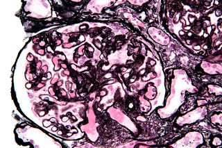
Membranous nephropathy is among the most common causes of the nephrotic syndrome in non-diabetic adult. It is an immune complex mediated disease which occurs when circulating antibodies permeate the glomerular basement membrane and form immune complexes with epitopes on podocyte membranes. Complement activation occurs and results in sublytic levels of C5b-C9 complexes resulting in release of oxidants and proteases that damage the underlying GBM.
Although the majority of MN falls under the idiopathic category ~ 75%, it is important to eliminate secondary causes where relevant as this distinction has important clinical implications; treatment of secondary nephropathy relies on treatment of the underlying disease process whereas idiopathic MN is treated using steroids and/or cytotoxic agents depending on risk of progression.
PLA2R is expressed on the cell body of podocytes near the foot processes. It undergoes constitutive endocytic recycling at the plasma membrane, providing a constant source of PLA2R. Upon binding of PLA2R antibodies, immune complexes aggregate and are shed into the subepithelial space. The identification of M-type PLA2R as the major target antigen in idiopathic membranous nephropathy in adults by Beck et al represented a paradigm shift in the diagnosis of MN. 70-80% of their patient population with idiopathic MN, but not those with secondary MN or controls, had circulating anti-PLA2R autoantibodies with a specificity is close to 90%–95% (see RFN coverage).
Staining of kidney biopsy specimens either by immunofluorescence or immunohistochemistry provides an assay by which to identify PLA2R associated MN. Detection of PLA2R in subepithelial deposits in kidney tissue is both a sensitive (69 to 84% across various studies) and specific (close to 100%) technique to diagnose MN. The rate of concordance between tissue PLA2R testing and serological PLA2R testing is variable among studies. One study showed 98% of tissue positive PLA2R patients were also seropositive. Other studies have shown less concordance and have generally found tissue testing more sensitive than serologic testing. Tissue deposits may persist even after serum antibody levels decline and it has been shown that 74% of PLA2R tissue positive patients were seropositive if the sample was taking during a period of heavy proteinuria as opposed to only 32% of patients being seropositive if samples were taken at time of complete or partial remission. Tissue testing is of particular relevance early in the disease course; at this stage circulating anti PLA2R antibodies may not be detectable because the kidney acts as a “sink”, absorbing antibodies. In these instances, PLA2R will be detectable on immunofluorescence staining but serum antibodies will be negative. With disease progression, renal tissue becomes saturated with anti-PLA2R antibodies and becomes seropositive and therefore can be used as a diagnostic and serological biomarker. As patients enter immunologic remission, anti-PLA2R remits in the circulation however proteinuria persists and it is likely that these patients would remain tissue positive if a renal biopsy were to be repeated.
Much research focus has shifted to serum anti-PLA2R antibodies and their diagnostic and putative roles as a biomarker of disease activity in primary MN. There are two standardised test systems for the qualitative and quantitative detection of anti-PLA2R autoantibodies: Anti-PLA2R IIFT and Anti-PLA2R ELISA. Studies a have shown a relationship between the presence and level of anti-PLA2R antibodies and disease activity. Immunologic remission usually precedes clinical remission by several months. This has particularly important treatment implications in that monitoring of PLA2R titres may help to identify those likely to achieve spontaneous remission and enable physicians to avoid immunosuppression. Antibody titres may also be important for prognostication at initial diagnosis.
- Hofstra et al found that patients with high antibody titre at time of diagnosis were less likely to achieve spontaneous remission. In a further study, the group determined that when patients were commenced on immunosuppressive treatment, the antibody titre falls rapidly, preceding the fall in proteinuria and concluded that the chance of achieving remission is higher if the initial anti-PLA2R antibody titre is low.
- Qin et al postulated that anti-PLA2R titre may have prognostic significance as there was a shorter time to clinical and biochemical remission in patients with low antibody titres. Levels at the end of treatment may also be beneficial in predicting long-term clinical outcomes.
- Bech et al found that almost 2/3 of patients with undetectable anti-PLA2R antibodies at the end of treatment in clinical remission, while all who had detectable anti-PLA2R antibody after therapy experienced clinical relapse.
In the time since PLA2R was reported as the specific podocyte antigen in primary MN, testing has become a standard part of the diagnosis and workup of MN. Assessment of both circulating PLA2R autoantibody and PLA2R in biopsy samples will likely have significant prognostic and therapeutic implications. The discovery of a reliable indicator of primary MN is particularly relevant given the “rule of thirds” and variable disease course.
Post by Laura Slattery, NSMC Intern 2018


