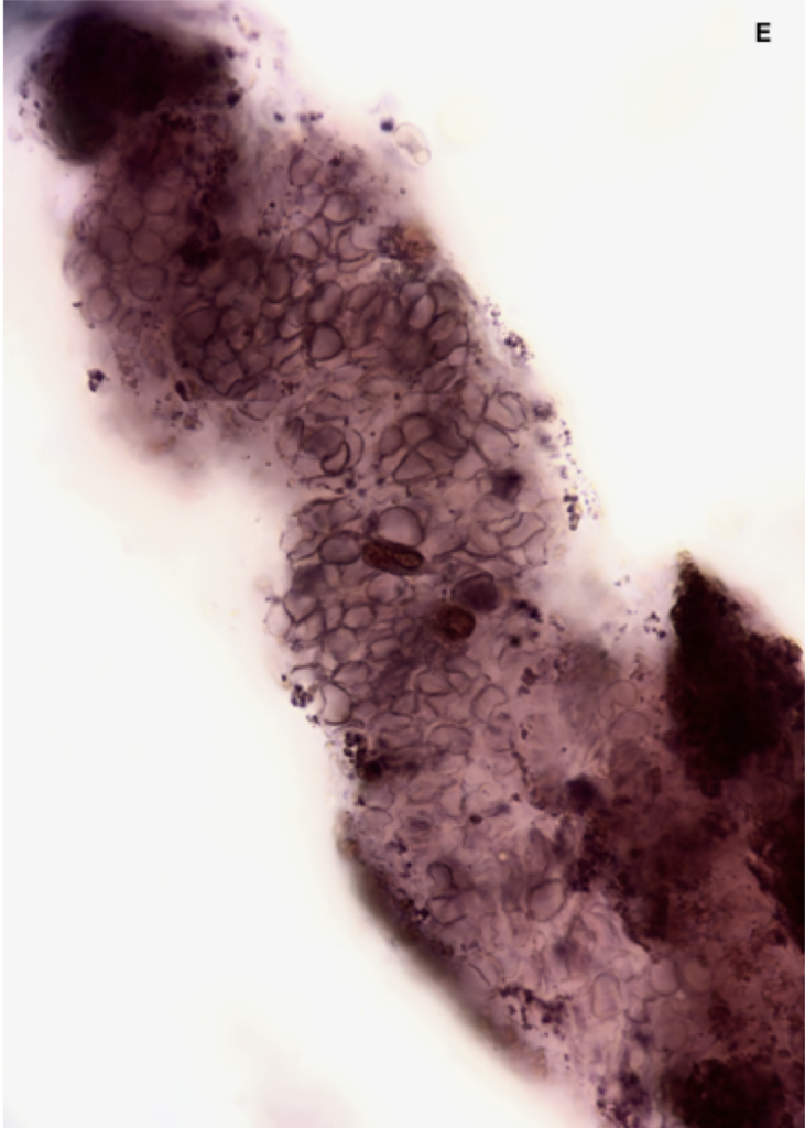
Red Blood Cell (RBC) Casts:
RBC casts (Fig. 1) are an important finding in the urinary sediment and most often signify the presence of a proliferative glomerulonephritis or vasculitic process. Although less common, RBC casts may also be seen in cases of acute interstitial nephritis, renal infarction, and in diabetic nephropathy.

Figure 1. RBC Casts A. BFI, SM stain; B. BFI, unstained C. Phase contrast, unstained D. BFI, with embedded renal tubular epithelial cells (RBC: red blood cell; BFI: brightfield illumination; SM: Sternheimer-Malbin)
RBCs may enter the tubular lumen via damaged glomerular capillary membranes or damaged tubular basement membranes. In acute interstitial nephritis, inflammation may lead to disruption of interstitial vessels and allow extravasation of RBCs into the tubular lumen via gaps in the tubular basement membrane. Hematuria and RBC casts in diabetic nephropathy may occur when areas of aneurysmal dilatation in glomerular capillaries rupture. This results in glomerular hematuria with little mechanical damage to the erythrocytes. In these cases, there may be RBC casts, but few acanthocytes, if any.
RBC casts form when RBCs which have entered the tubular lumen are trapped within solidified Tamm-Horsfall protein matrix. These casts may have a variable appearance with some containing only a few RBCs and others so densely packed that individual cells and protein matrix are difficult to see without higher magnification.
RBC Degeneration, Ghost Cells, & Hemoglobin Casts:
When RBC casts initially form, the cells retain their shape and hemoglobin content. As they begin to degenerate, their appearance changes considerably. As RBCs become “depigmented” they appear as “ghost cells” -the cell outline is visible, but the cytoplasm and pigment are no longer apparent (Fig. 2). During the degeneration process denatured hemoglobin forms “Heinz bodies” which appear as small dense granules along the cytoplasmic membrane (Fig. 2). The heme released in this degeneration process pigments the cast a yellow/orange or reddish/brown color and at this stage, when the cast is pigmented without obvious cellular remnants, it is termed a hemoglobin cast (Fig. 2).
Degenerating RBCs, SM stain ![]()
Various stages of degenerating RBCs with Heinz bodies (denatured Hgb clumps) 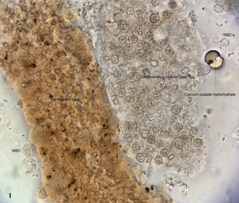
(R) Degenerating RBCs with Heinz bodies (L) Hgb cast ![]()
Intact and degenerating RBCs with pigmented granular material (Hgb) 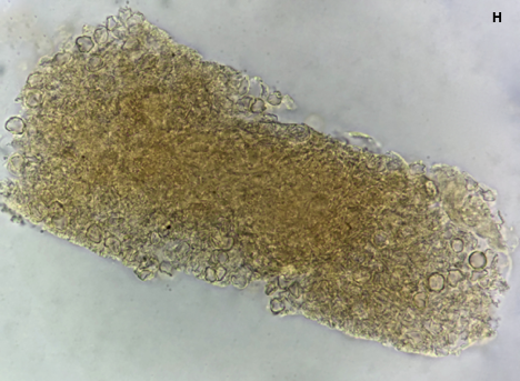
Hgb cast, due to progressive RBCs degeneration
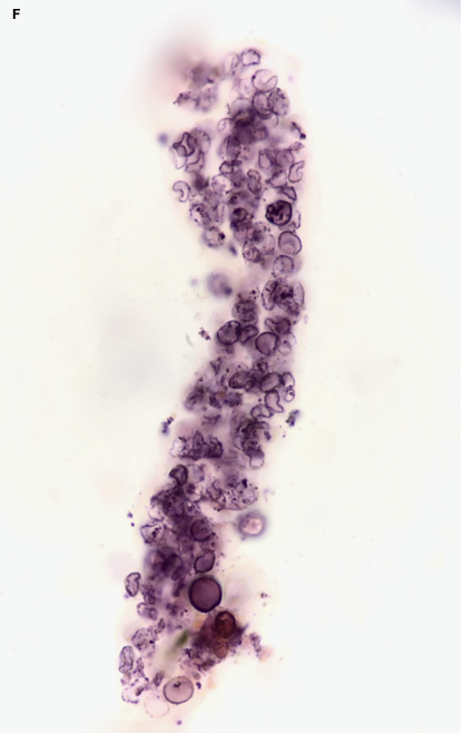

Figure 2. RBC casts with degenerating RBC, hemoglobin, and Heinz bodies. Click on the individual images to expand. (RBC: red blood cell; Hgb: hemoglobin; SM: Steinheimer-Malbin; BFI: brightfield illumination; R: right; L: left)
Red blood cell casts can be an elusive finding and require a diligent search, often using different illumination modalities and magnifications. What appears on low power to be a dense granular cast may upon review under higher magnification actually contain RBCs! RBC casts are fragile and easily degenerate. They are subject to fragmentation or disruption at higher centrifuge speed and may disintegrate if the sample is left too long before examination.
Microscopy tip: remember that while phase contrast provides enhanced contrast of elements with a low refractive index, it does so at the expense of resolution. In many cases, brightfield illumination provides the best resolution, especially of pigmented or stained specimens. When it is difficult to determine the type of cell or cast you are viewing, try the oil immersion lens – this increases resolving power by increasing the numerical aperture of the objective lens and can assist in identification and interpretation.
Post by: Jay Seltzer, MD