After centrifugation of the urine specimen, the macroscopic appearance of the sedimentation can offer certain clues about the nature of the urinary sediment.
Color is by far the most important characteristic to define different sedimentations, but other features such as diffusibility, translucency and adhesiveness have been used. In this post I will consider only the following categories, which can be distinguished rather reliably:
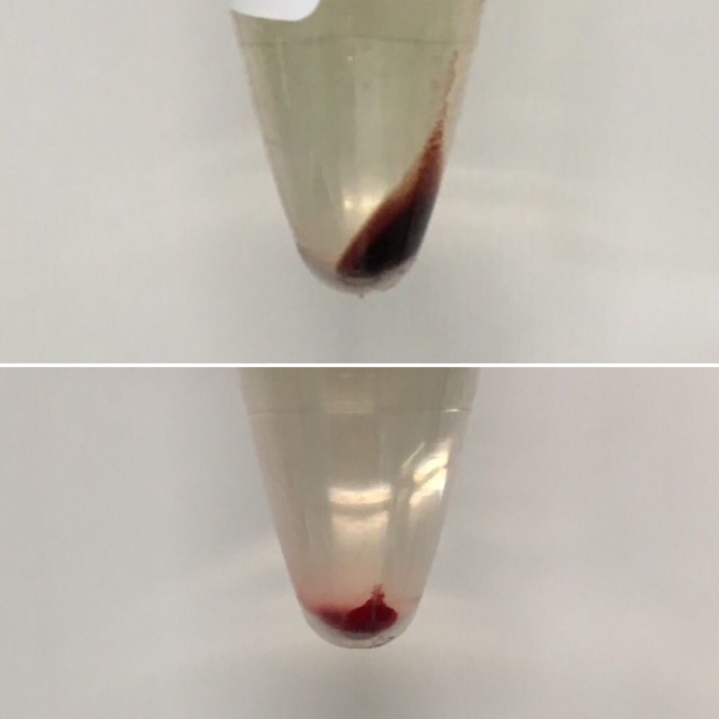
1. Dark or bright Red (bloody)
2. Brick-red or Reddish-brown
3. White
4. Cloudy
1. Dark or bright Red (bloody)
Blood in the sediment is usually easily recognized by its dark or bright red color (Fig. 1). Generally, erythrocytes are found at the bottom in mixed sediments.
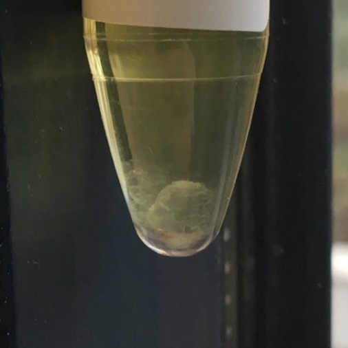
2. Brick-red or Reddish-brown
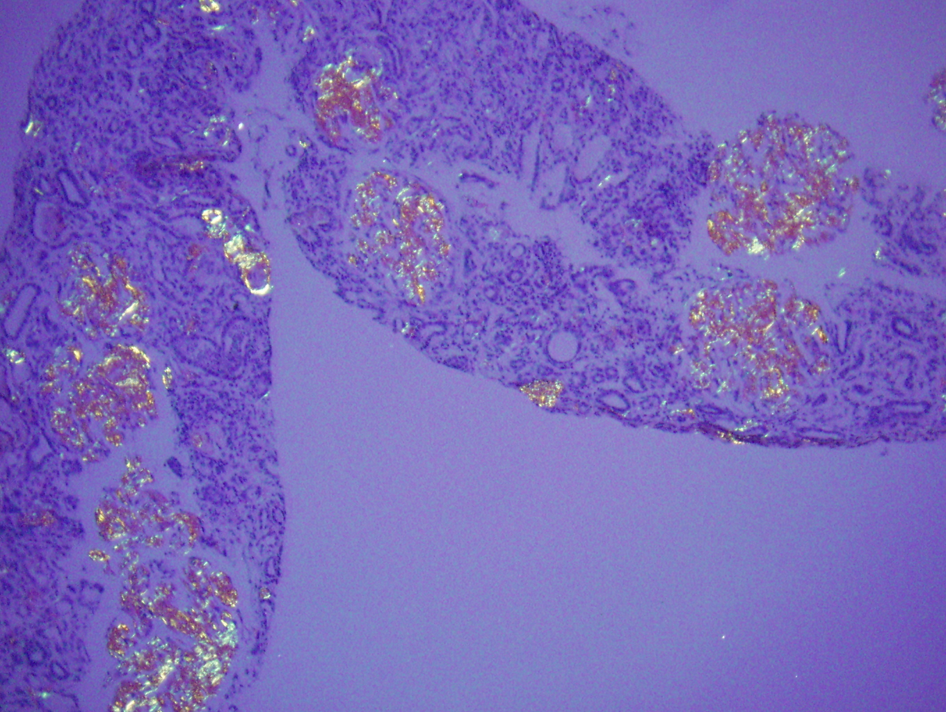
A brick colored sediment, the so called sedimentum lateritium (Fig. 2) is typical for high amounts of amorphous urates. Even in much more brownish sediments of urates a reddish-orange hue is readily apparent. Resuspension reveals a grainy structure (Fig. 3).
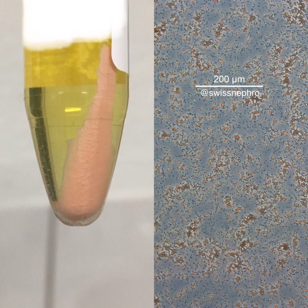
amorphous urates. (Right: Phase contrast, original magnification x100)

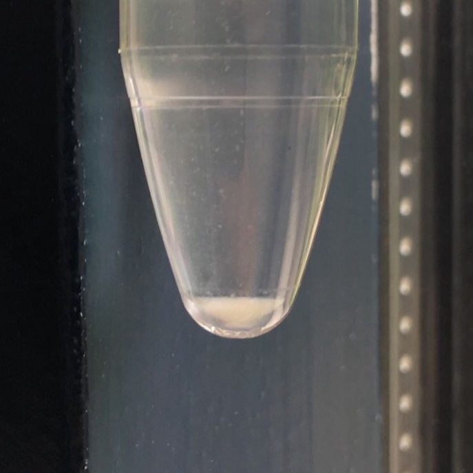
3. White
Amorphous phosphates, leucocytes and vaginal fluor can all give rise to predominantly white sediments. Whereas leucocyturia produces a creamy appearance and yellowish tinge (Fig. 4), amorphous phosphates present with bright white coloration (Fig. 5). Vaginal contamination is oftentimes more grayish (Fig. 6).
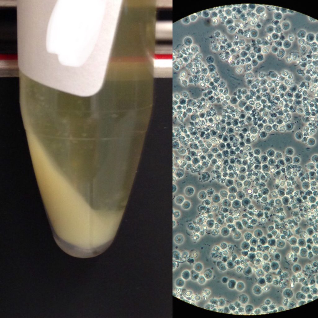
Phase contrast, original magnification x400)
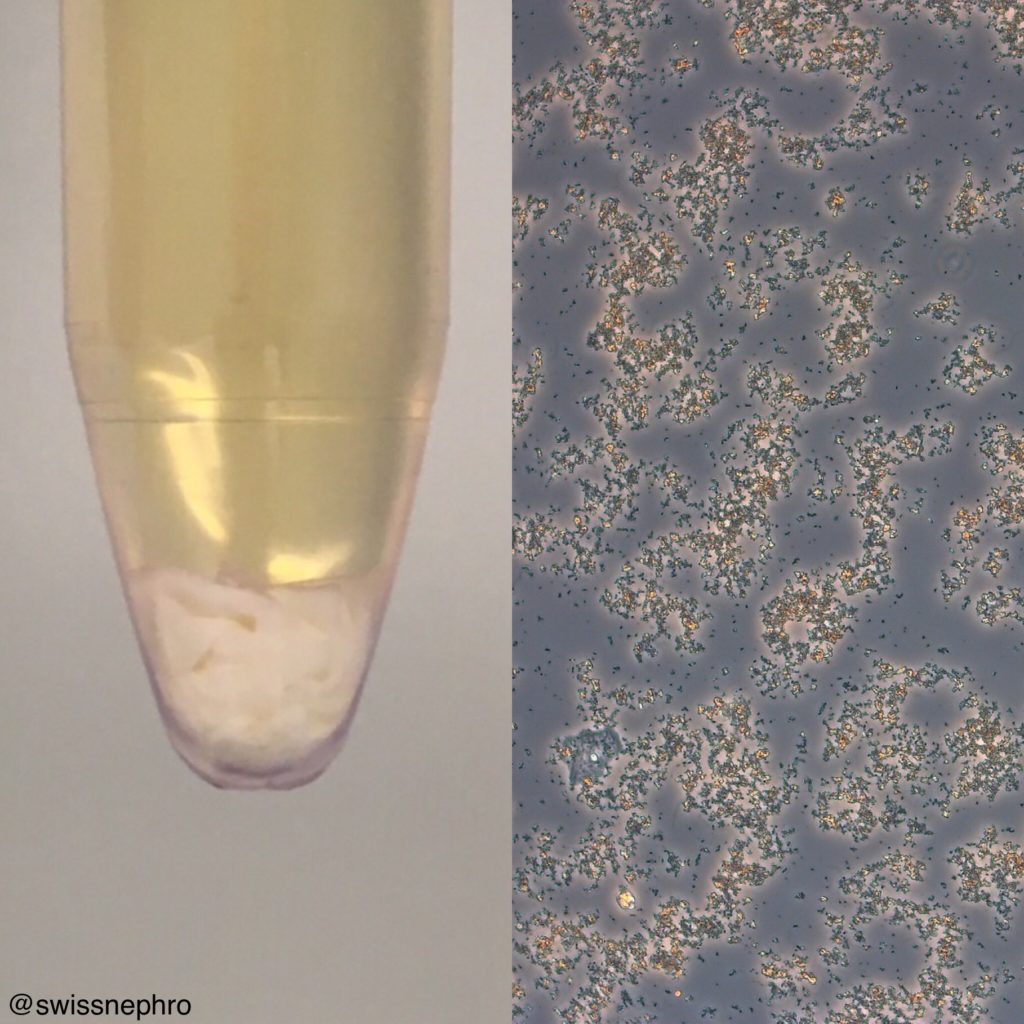
original magnification x100)
4. Cloudy
Mucus can form rather loose, more or less free-floating clouds at the bottom of the tube. (Fig. 7).
Macroscopic sediment inspection is not rocket science and its clinical relevance is limited. I use it as a quick global assessment of the sediment, to grasp the degree of hematuria and pyuria, and in the differentiation of amorphous phosphates and urates.
Further Reading
Rieder H. Macroscopic characters of sediments. Pages 5-7 in Rieder H, Atlas of urinary sediments. London 1899.
Post by: Florian Buchkremer

white half spheres in clear pale yellow urine? The half spheres have sharp contours… never seen anything like that…
Thank you for this article, it really helped me differentiate what kind of macroscopic sediment I have in urine. I was searching Google all way through for like 20 minutes but still did not find a satisfying image resembling to my sediment. Then I found your article and found out it was very probably many pieces of mucus. I’m about ovulation now so I guess it can have some connection. Thank you.
I have all these types of urine at differing times..can have all in one week.or reacuring for months at a time. Except the first one..I don’t get that.
I would share them on Twitter
How can I attach the pics?
I have several pics of urine. Can you help?