Damage to the glomerular basement membrane allows plasma lipids to enter the urinary space. These lipids may appear in various forms, either as free lipid droplets, lipid casts, oval fat bodies, and/or as cholesterol crystals. In most cases, this process occurs in the setting of nephrotic proteinuria, however lipiduria may also be present in polycystic kidney disease and Fabry Disease.
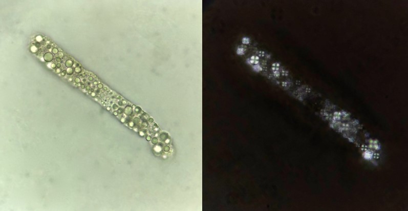
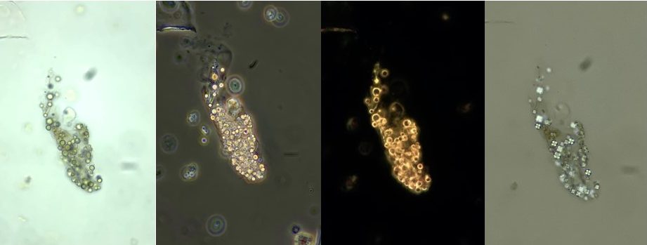
Lipids that enter the tubule are to some extent absorbed by kidney tubular epithelial cells (and macrophages). The intracellular lipids coalesce into refractile globules within the tubular cells forming what are called oval fat bodies (Figure 4). As these lipid engorged tubular cells slough from the tubule they appear in the urine either free, or within casts Figure 1, 6). Lipid casts are the result of accumulated free lipid droplets or lipids released from degenerating oval fat bodies. Although less common, cholesterol crystals may form which appear as large notched geometric plates (more likely to be seen in refrigerated specimens).
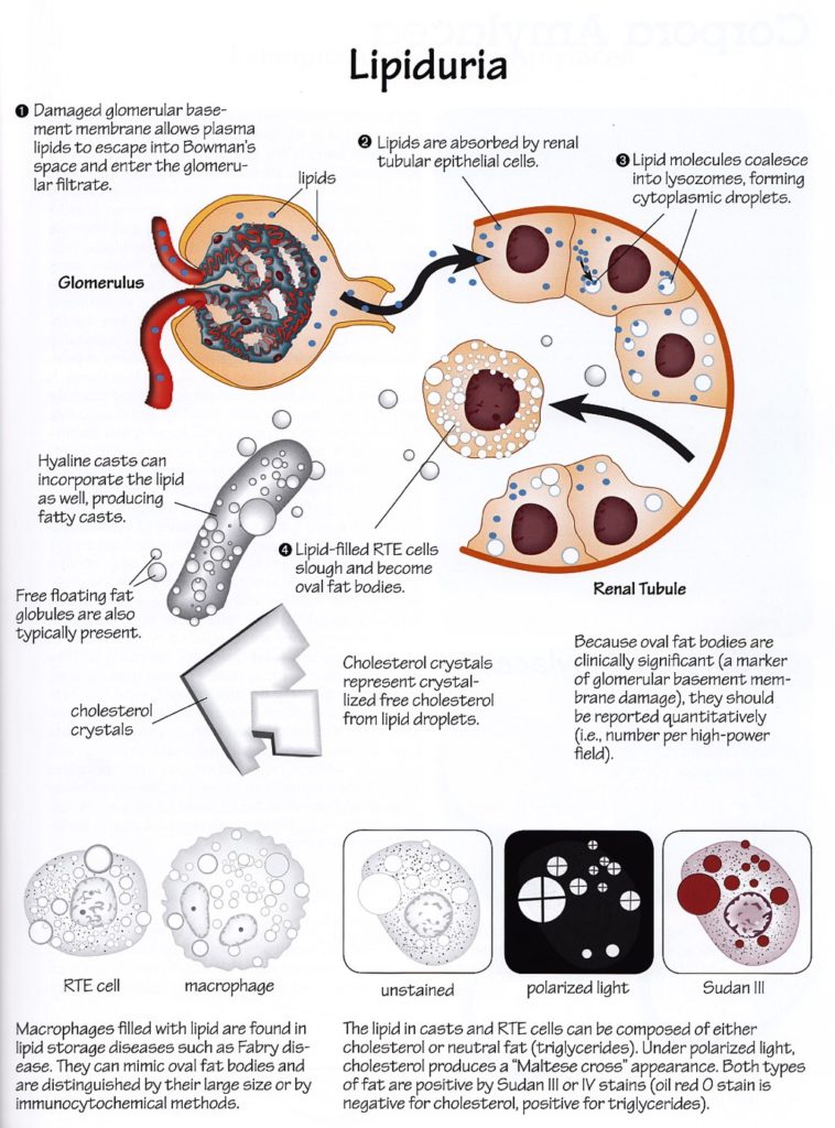
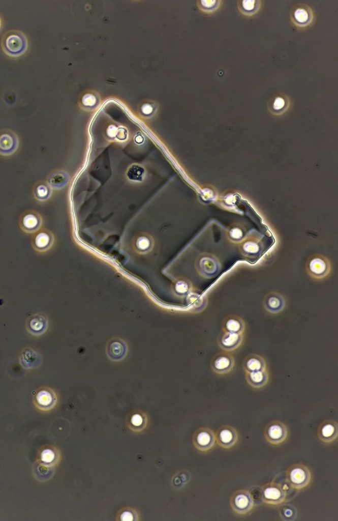
Cholesterol crystals (Figure 3) demonstrate a colorless birefringence unlike the polychromatic appearance of radiologic dyes which may otherwise appear similar.
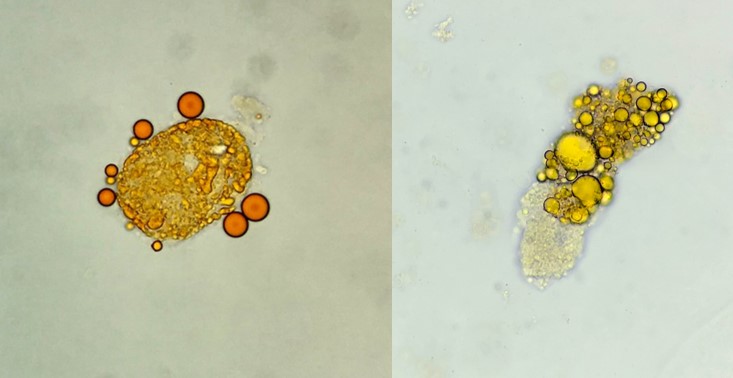
Lipids droplets are round, refractile, and of variable size, ranging from much smaller than an RBC to larger than a tubular epithelial cell. They are best appreciated under darkfield illumination (because they are highly refractile) and under polarized light which reveals a characteristic “Maltese cross” pattern of birefringence (Figure 1 and 4).
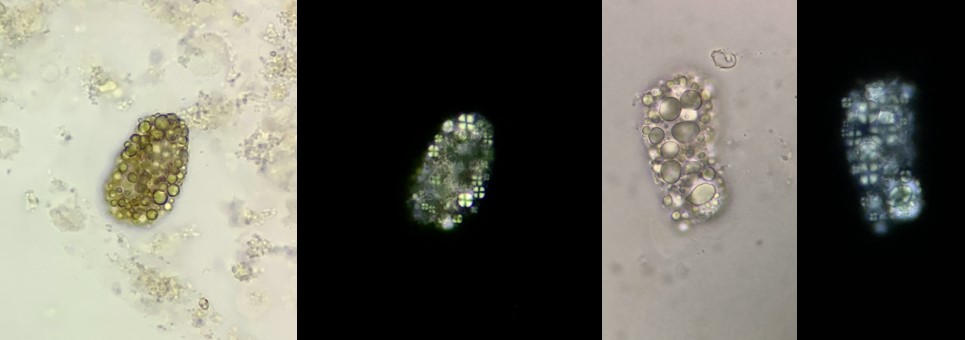
Remember, however, that not all types of lipid display this “Maltese cross” birefringence. This pattern is characteristic of cholesterols and may not be seen with neutral fats. In such cases, these lipids can be readily identified by staining with Sudan III or Oil Red O, which are lysochrome diazo dyes which stain neutral triglycerides and lipids (Figure 5).