If you’re regular reader of Urine Sediment posts, you know all about red cells, white cells, and epithelial cells by now – but what about the zebras of the urine sediment? In 2019 we talked about the so-called “decoy cell” that can identify Polyomavirus BK infection in allograft recipients. Today, we’ll cover a few other uncommonly encountered cells in the urine and what they mean clinically.
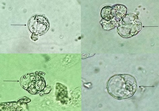
Bubble cells. These are a real unicorn and have only been described in the literature on one occasion. “These cells were bizarre, large cells with a single nucleus, which appeared to contain one or more fluid-filled vesicles.” Bubble cells were most prevalent in the sediment of patients with acute tubular necrosis but were also seen a variety of other renal diseases. In most patients with acute tubular necrosis, the sediment also contained “normal”-appearing renal tubular cells, muddy brown casts, and oval fat bodies which were indistinguishable from those seen in the nephrotic syndrome. By electron microscopy, the bubble cells appeared to be vacuolated renal tubular epithelial cells, which had characteristics of viable cells.”
Though not spotted often in literature, bubble cells themselves are not extremely rare. The cells observed in Figure 1, for example, are from a patient with nephrotic range proteinuria and severe tubular and glomerular damage due to focal and segmental glomerulosclerosis.
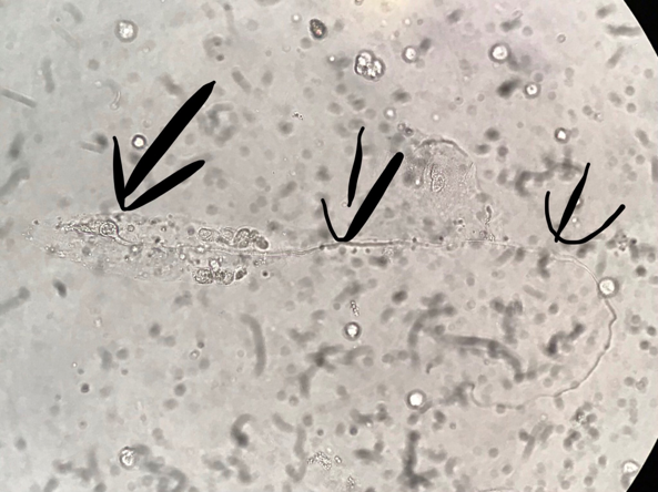
Tadpole cells or snake cells are elongated cells where the nucleus stays visible on one side of the cell structure and a long “tail”-like structure is seen on the other. This kind of cell can be observed on the urine sediment of patients with malignant keratinizing squamous cells originating from a tumor in the urinary system. Expect to see these cells much like you would leukocytes and urologic hematuria in the setting of tumors in the urinary tract.
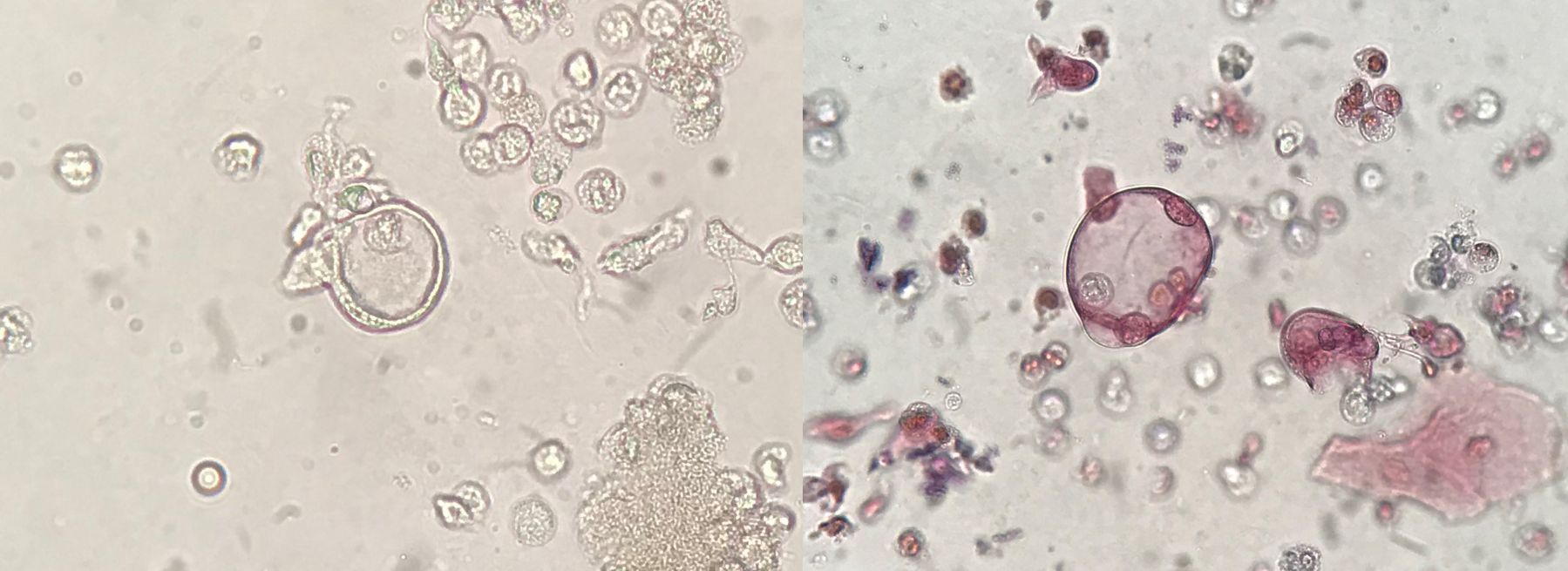
Signet ring carcinoma is an exceedingly rare and aggressive variant of primary bladder carcinoma. The cells of the tumor can be seen in urine, but cytomorphologic features in urinary specimens have not been well characterized. These cells can be easily missed or misinterpreted due to their rarity and singly dispersed nature.
Here we review three examples of unusual and abnormal cells that can be observed in the urine sediment. Morphological modifications of the nucleus and of the cytoplasm of the cells, intranuclear inclusions and intracytoplasmic inclusions can help with diagnosis of important clinical conditions such as malignancy or tubular necrosis. While fresh and unstained urine sediment analysis does not replace urine cytology on the diagnosis of tumors, it can be a tool to aid diagnosis or prompt further investigation.
So remember to keep these less common cell types in the back of your mind when you examine urine sediment- as DH Lawrence said: “The eye doesn’t see what the mind doesn’t know”!
Post by José A. T. Poloni

Muchas gracias por la información.