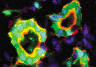
 The study of AKI/ATN has relied heavily on one particular animal model: the warm ischemia-reflow model (often referred to as “ischemia-reperfusion injury”), in which one of the renal arteries is transiently ligated off for a set period of time while body temperature is maintained, then opened up and allowed to reperfuse the kidney. A recent review in Kidney International by Heyman et al addresses some of the limitations of this model, mostly in terms of differences between the mouse model of ischemia-reperfusion and the typical human AKI/ATN we experience clinically.
Some the important differences between mouse and human AKI: First, while the warm ischemia-reperfusion model tends to initially target the S3 segment of the proximal tubule and typically leads to overt tubular necrosis, in human AKI necrosis is not always present, and when it is tends to be patchy and most commonly affecting the distal nephron (in particular: the medullary thick ascending limb and medullary collecting ducts). Second, in clinical practice it is COLD ischemia which is often the mechanism of injury (e.g., surgical procedures in which the aorta is cross-clamped is often performed in the setting of a lowered core temperature, and donated kidneys are typically stored on ice prior to transplantation), rather than warm ischemia. Overall, the authors conceded that the rodent ischemia-reperfusion model has been invaluable, but caution against using it to explain all aspects of human AKI/ATN.
The notion of whether AKI/ATN occurs primarily in the proximal versus the distal tubule is not merely of academic interest. According to one version of the “distal nephron model”, the reduced GFR experienced in response to medullary hypoxia is actually an adaptive response: decreasing the metabolic demands of tubular epithelia would decrease hypoxic injury; in a sense one could think of the nephrons as “hibernating” in a low metabolic state until they sense that the hypoxic insult has been removed and they can resume optimal cellular function. If this is true, it might caution physicians from trying to stimulate GFR in patients with AKI, instead encouraging attempts to limit tubular energy expenditure.


Nice post.Really thought provoking for researchers!
Thanks…