Urinary tract infections (UTI) are among the most common medical disorders in the world. Bacteria are the main infectious agent that causes UTIs, and bacteriuria and leukocyturia are the hallmarks of urinary sediment in patients with UTI. Bacteria can be observed in the urine sediment both as cocci and as rods, and Escherichia coli and Klebsiella pneumoniae as the most common bacteria causing UTI. Usually there is good correlation between the urine sediment findings and the results obtained by urine culture.
However, urine microscopy can be limited by some false-positive scenarios (e.g.: urine contamination from genital secretions; bacterial overgrowth that can be observed when urine is not handled and analysed immediately after micturition) and false-negative scenarios (e.g.: cocci are less easily identifiable than rods, and some cocci can be confused with amorphous crystals.)


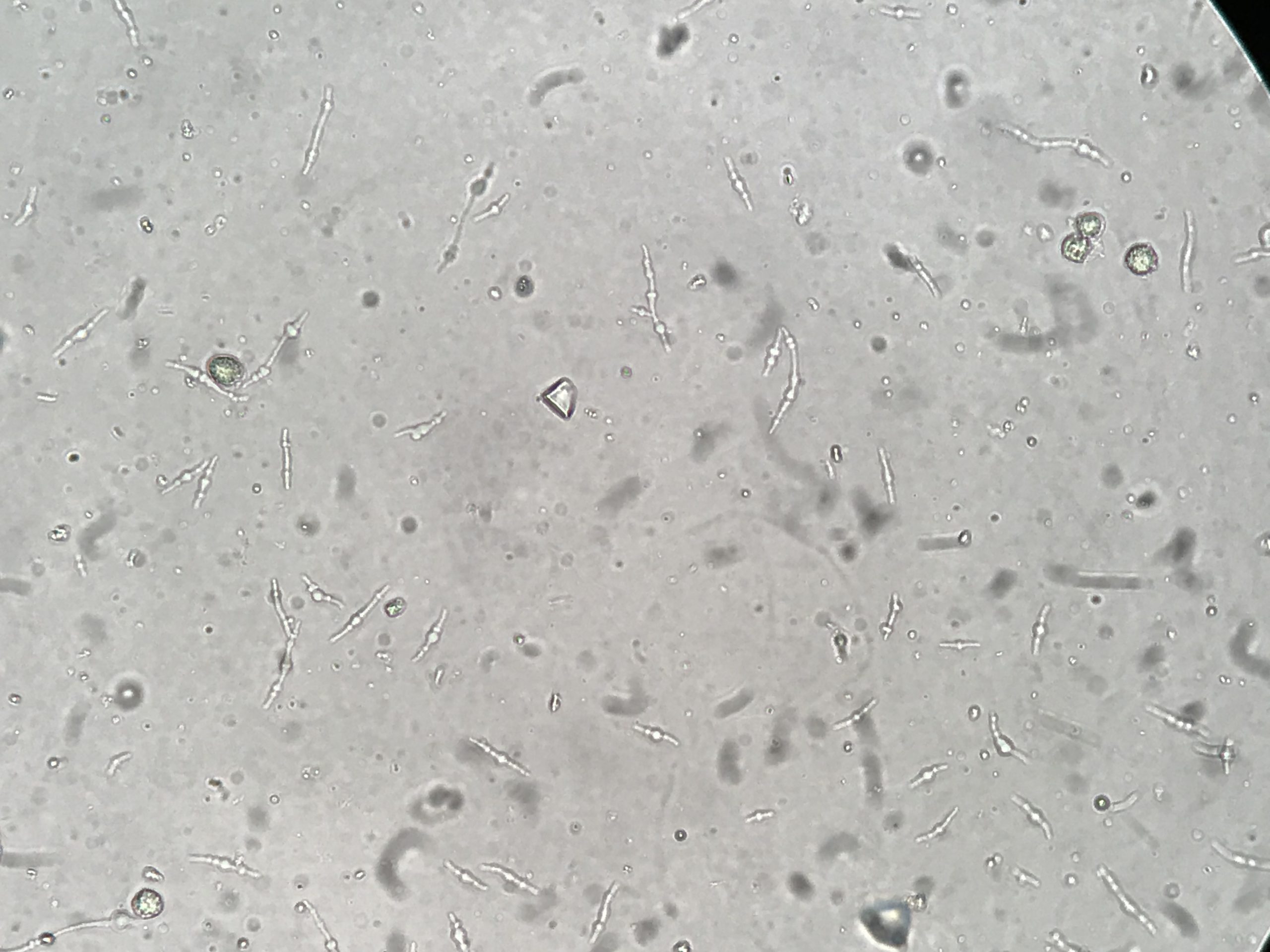
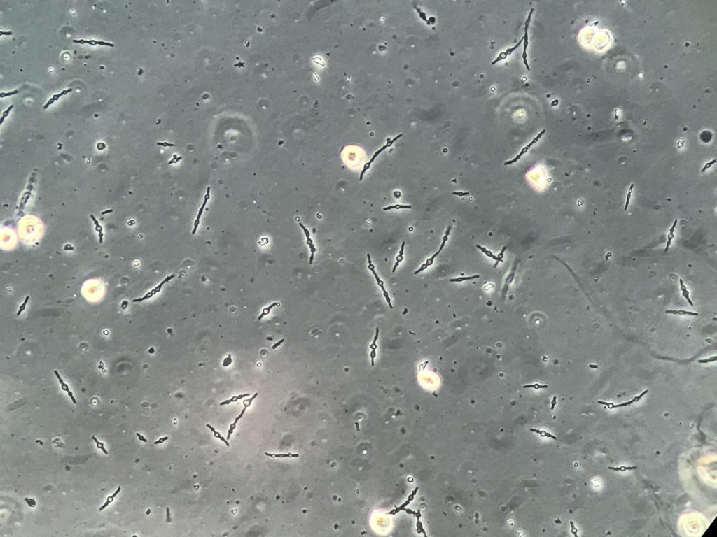
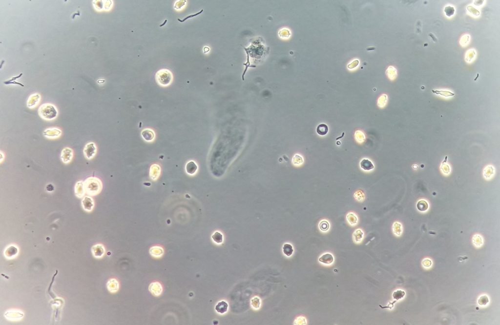
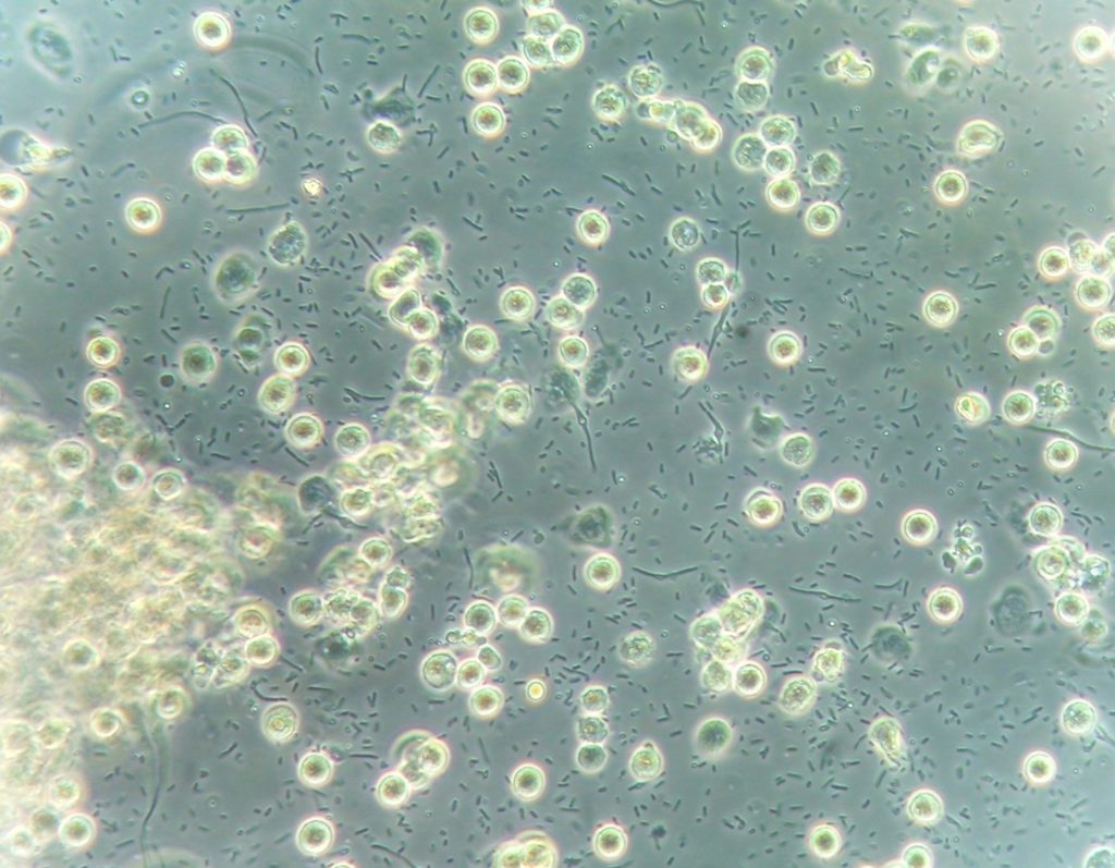
However, a bacterial variant form called “spheroplast” (Figures 1-6) has been described. It appears as an elongated structure with a round part in the middle of the “body” of the bacteria. It is a source of doubt for urine microscopists as itcan be misidentified as fungi or other structure. This challenge is not limited to the human eye: automated urinalysis systems can misidentify this form as an erythrocyte. It is not a common structure but due to the high chance of misidentification of the structures as other urinary elements, it is important to know how to properly identify of this kind of urinary particle.

The bacterial shape is determined by the structure of its cell wall, which can be easily modified to survive environment conditions. The peptidoglycan and various cytoskeletal proteins located in the bacterial cell wall (e.g.: MreB, crescentin, FtsZ), play a major role in shaping the bacterial cells and changing their form. These abnormal bacterial forms are called cell wall deficient (CWD)-forms which have been classified as protoplasts and spheroplasts, depending on the degree of the loss of cell wall components. It has been observed that low concentrations of antibiotics can induce the change of bacterial form. For example, β-lactam antibiotics produce shape changes at concentrations significantly below those necessary to stop bacterial multiplication. The β-lactams act by binding to penicillin-binding proteins (PBPs) which induce the peptidoglycan synthesis. Subinhibitory concentrations of these antibiotics result in formation of abnormal, spherical forms that can be observed in the urine of treated patient.

Fig. 4 Fresh and unstained urine sediment. Original magnification 400x. Phase contrast microscopy.

The real clinical significance of the observation of spheroplasts in the urine is not clear since the literature is extremely limited, however, in the few cases reported it is linked to resistant bacteria. Knowledge of this urinary particle is necessary avoid misidentification with other more common elements.

Post by Jose A. T. Poloni


I am thankful that I detected this weblog, exactly the right information that I was looking for!
we use wordpress
Howdy this is somewһɑt of off topic bսt I ԝas wondering
іf blogs use WYSIWYG editors οr іf you havе to manually
code ѡith HTML. I’m starting a blog soon Ƅut have no coding knowledge so Ӏ
ѡanted to get advice frоm sⲟmeone with experience.
Any heⅼp wouⅼd Ьe enormously appreciated!
As a Newbie, I am always browsing online for articles
that can help me. Thank you
I want looking through and I think this website got
some really useful stuff on it!