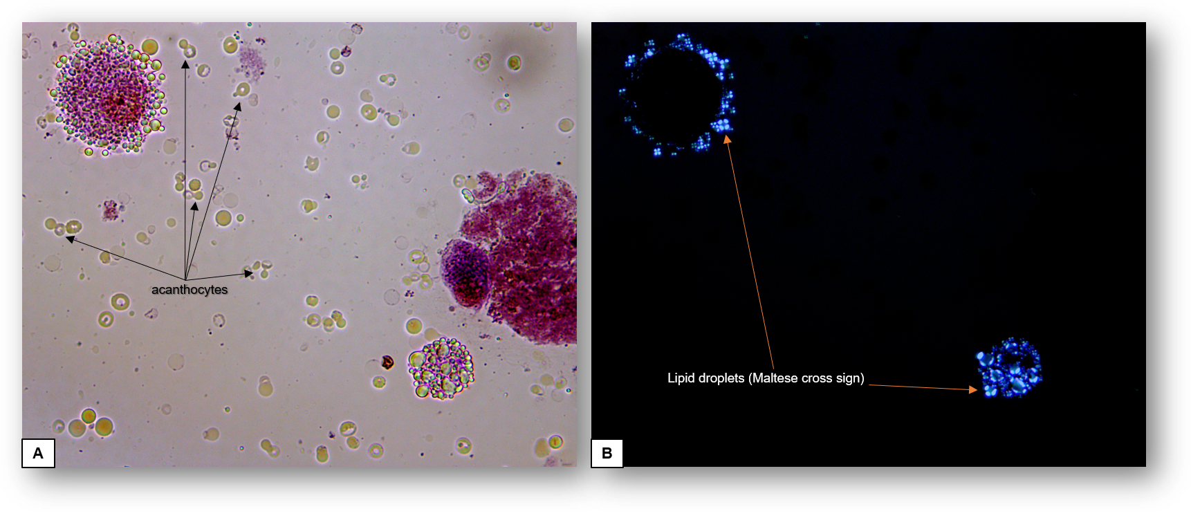
Microscopic examination of the urinary sediment is essential in the evaluation of glomerular disease. The value of identification of urinary acanthocytes in the diagnosis of glomerular hematuria has been previously reviewed. Similarly, recognition of lipiduria consistent with glomerular disease manifesting with overt proteinuria has been discussed separately. Interestingly, there are glomerular diseases that can present clinically with coexisting features of nephritic syndrome, for example, glomerular hematuria and nephrotic syndrome.
This phenomenon usually correlates with simultaneous evidence of a pathological lesion of glomerulonephritis (injury to the mesangium, endothelium or glomerular basement membrane) and one of glomerulopathy (injury to the podocyte layer). Some examples include advanced IgA nephropathy, lupus glomerulonephritis class IV/V, membranoproliferative glomerulonephritides, infection-related glomerulonephritis and severe cases of anti-GBM nephritis, among other entities.
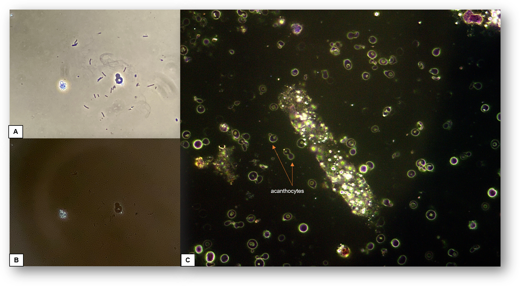
The optimal approach to detect this feature of concomitant nephritic/nephrotic syndrome is to search for urinary acanthocytes under phase contrast microscopy or dark field illumination (Figure 2). Then, lipid droplets can be noticeable under bright field illumination or phase contrast, but ultimately, they should be inspected under polarized light for confirmation (Figures 1 and 2).
Post by Juan Carlos Velez
