Anti-Nephrin Antibodies in Minimal Change Disease
Approach to the Patient with Peritoneal Dialysis Catheter Dysfunction- Part 1
Want to write a case report? RFN has you covered with a list of journals to choose from
Have a Nephrology Question? There Might Be A #Tweetorial For That!
Conferences, Courses, & Grants
FOAMed
Interventional Nephrology Series
Focus on Point-of-Care Ultrasound in Nephrology (POCUN) Series
Kidney Biopsy of the Month Series
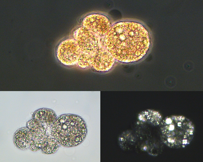
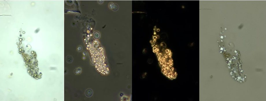
Urine Sediment of the Month: Isolated Lipiduria
What is “isolated lipiduria”? The detection of urinary lipid droplets with a typical Maltese cross appearance in the absence of proteinuria or albuminuria. There are no reports or systematic analyses of this phenomenon, so this post reflects anecdotal…
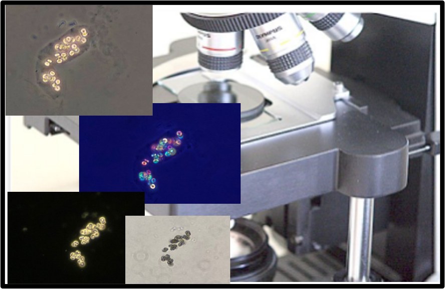
Urine Sediment of the Month: Transmitted Light Microscopy Techniques
Although it remains a polarizing topic, looking at urine has come a long way since the dark ages before wide availability of renal biopsy. Not just a passing phase, modern urine microscopy allows the bright nephrologist to readily…
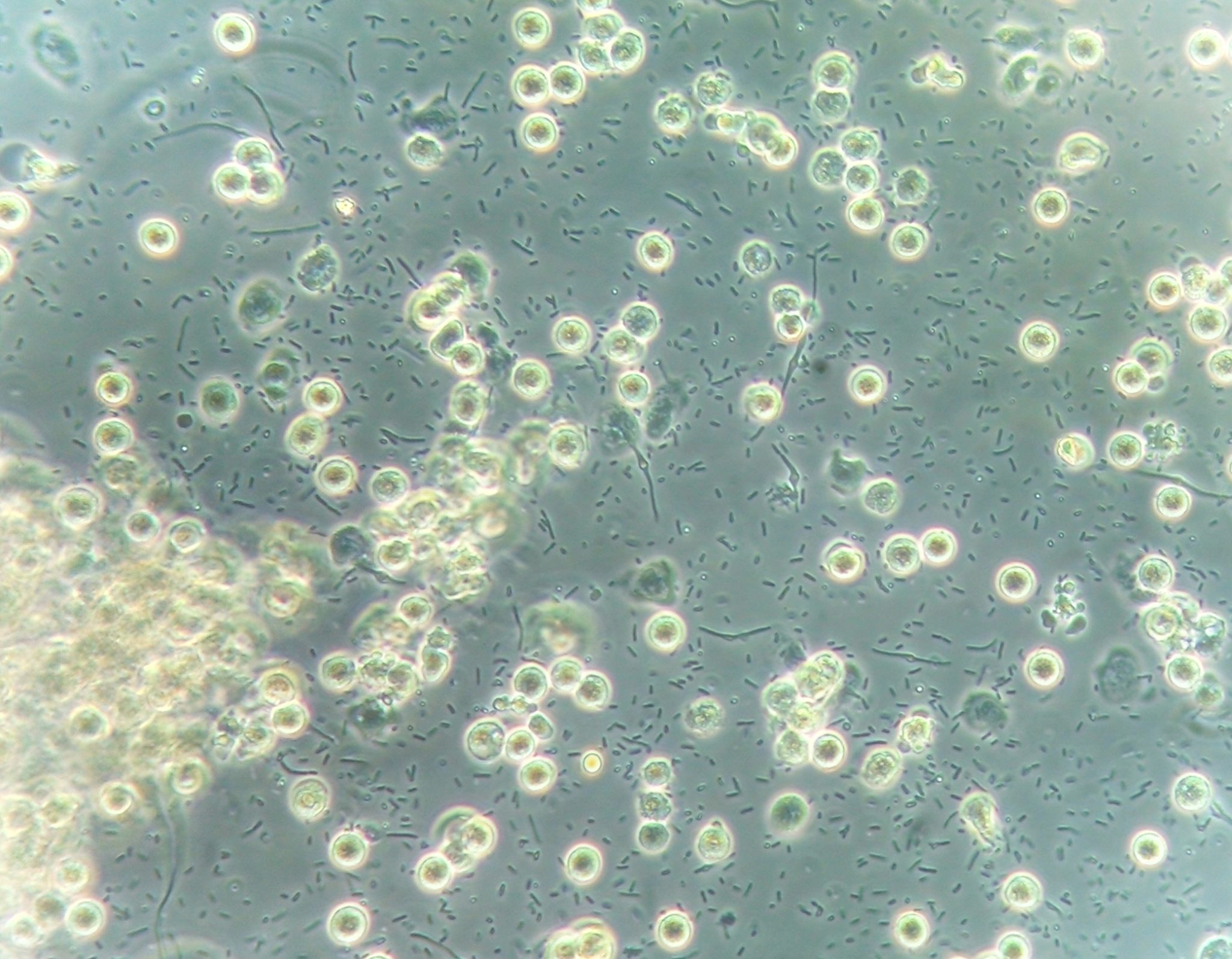
Urine Sediment of the Month: Bacterial Variant Forms
Urinary tract infections (UTI) are among the most common medical disorders in the world. Bacteria are the main infectious agent that causes UTIs, and bacteriuria and leukocyturia are the hallmarks of urinary sediment in patients with UTI. Bacteria…
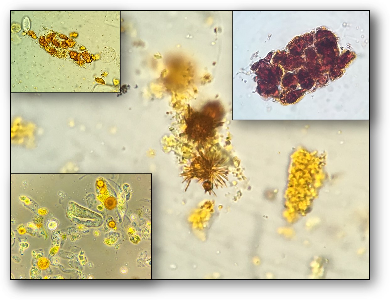
Urine Sediment of the Month: Findings in Cirrhosis, Cholestasis, and Hyperbilirubinuria
Microscopic examination of the urinary sediment in the context of hyperbilirubinemia and increased urinary bilirubin excretion requires special attention to the unique chromatic characteristics acquired by the specimens. As serum bilirubin increases, a yellow-tinged urine becomes increasingly noticeable….

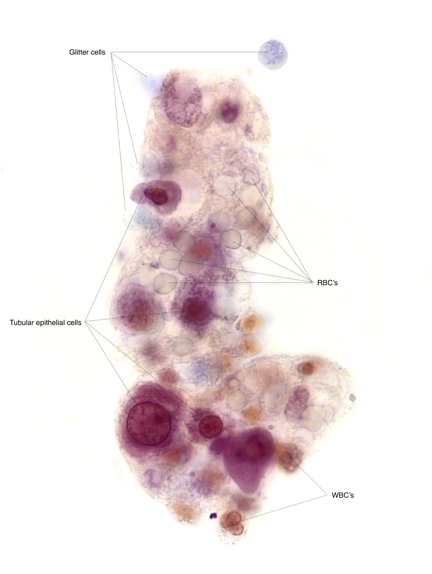
Urine Sediment of the Month: 4 Flavors of Nucleated Cells
In the routine urinary sediment examination, you may come across 4 types of nucleated cells: 1. Squamous epithelial cells2. Leukocytes3. Transitional epithelial cells4. Tubular epithelial cells Let’s discuss how to differentiate between them. Squamous epithelial cells are easily…
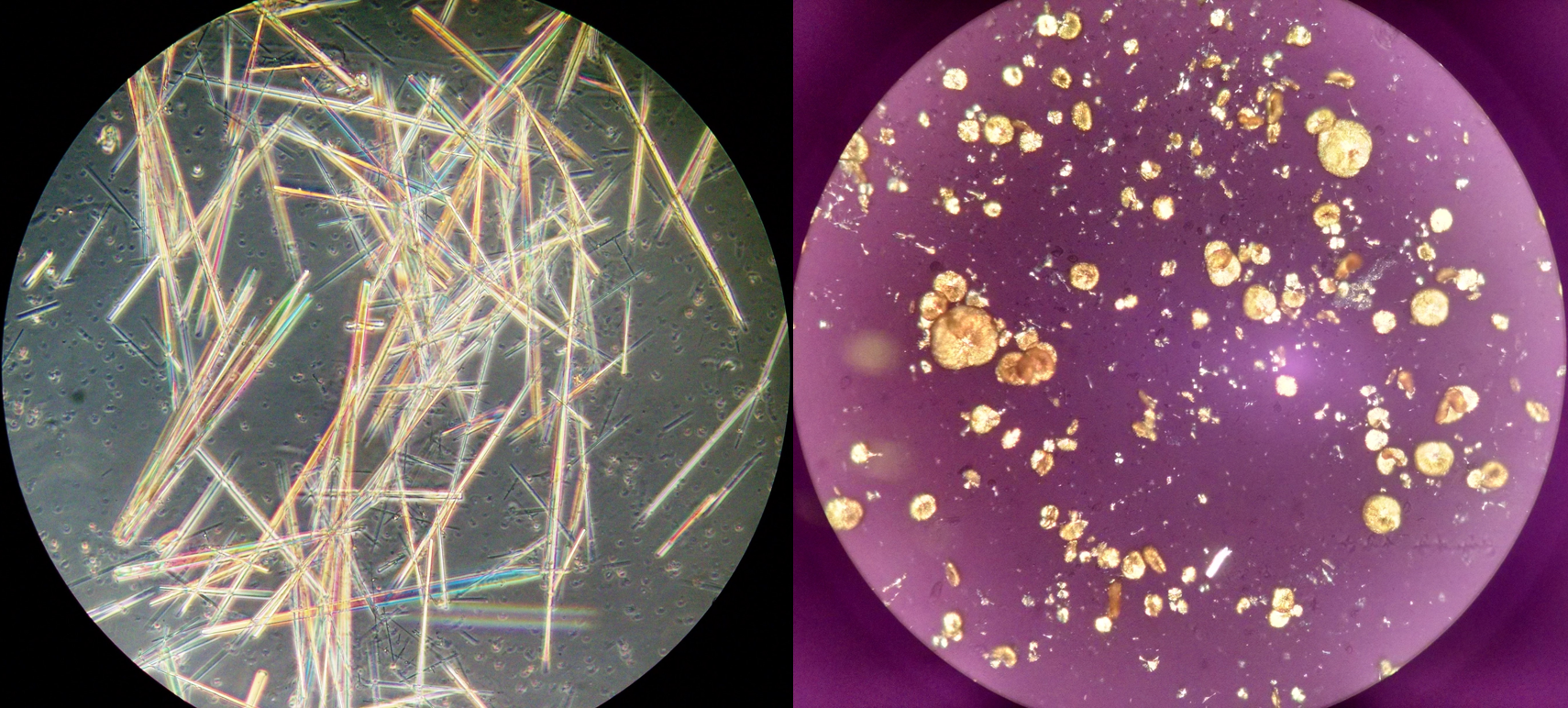
Urine Sediment of the Month: All About Those Oval Fat Bodies
Damage to the glomerular basement membrane allows plasma lipids to enter the urinary space. These lipids may appear in various forms, either as free lipid droplets, lipid casts, oval fat bodies, and/or as cholesterol crystals. In most cases,…
Urine Sediment of the Month: Drugs & Crystalluria
Several drugs, mainly antimicrobial or antiviral agents, can cause transient crystalluria, in isolation or in conjunction with other urinary abnormalities and a wide range of clinical implications. The factors favoring the formation of drug crystals are drug overdose,…
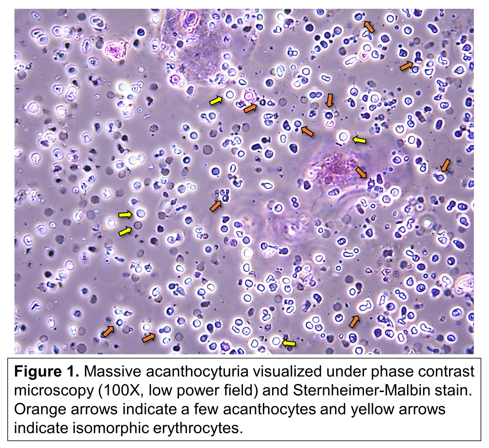
Urine Sediment of the Month: Dysmorphic Red Blood Cells, Blebs, & Spikes
Urine acanthocytes are a distinct type of dysmorphic erythrocytes that can be found during microscopic examination of the urinary sediment. Their unique characteristics make them rather pathognomonic of hematuria that originates from a glomerular disease, i.e., glomerular hematuria…
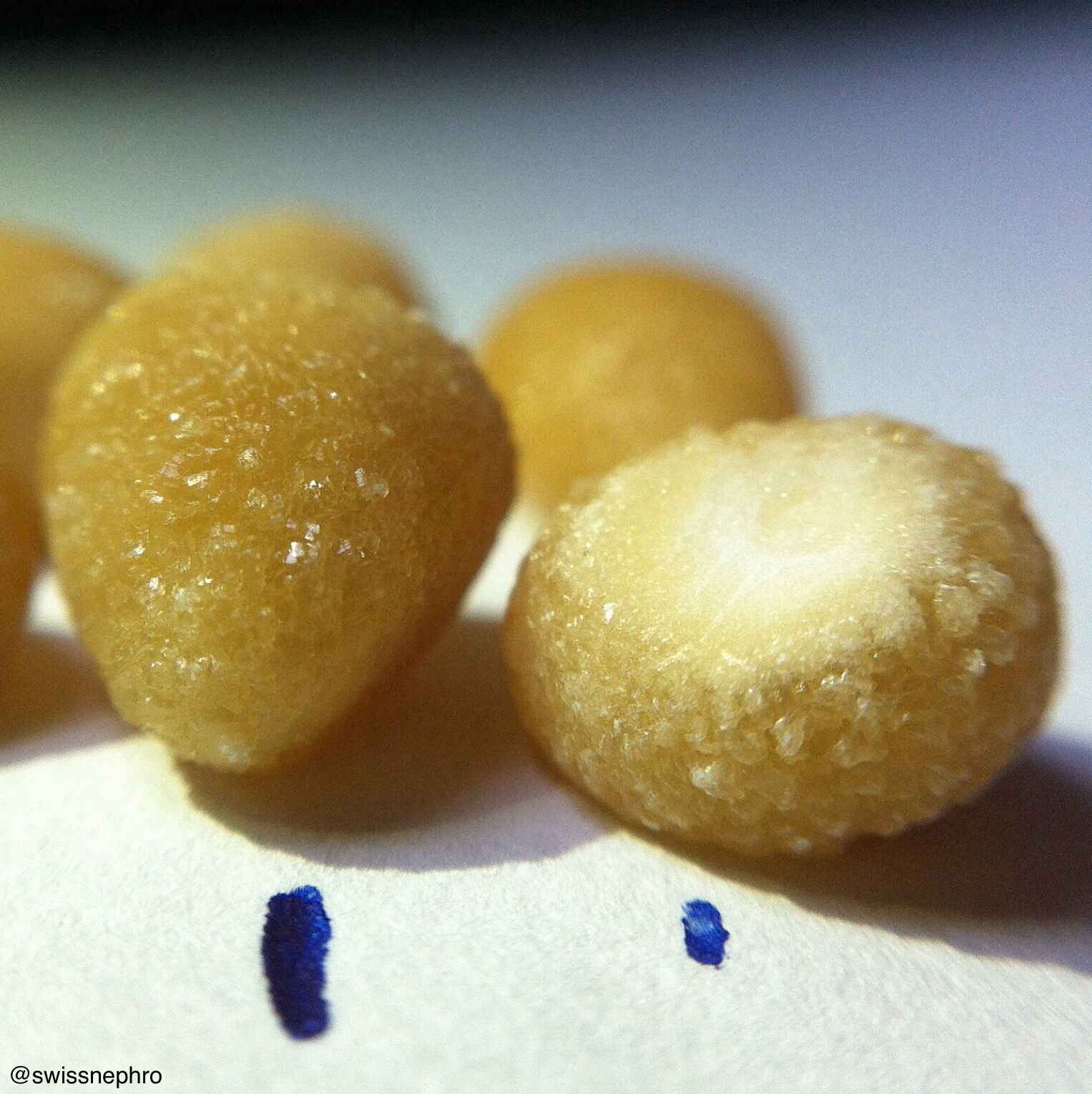
Urine Sediment of the Month: Cystine Crystals
Cystinuria is the most common kidney stone disease with Mendelian genetics. Caused by mutations in SLC7A9 and SLC3A1, affected patients excrete high amounts of cystine in their urine and are vexed by recurrent episodes of nephrolithiasis. Cystine stones…
Urine Sediment of the Month: A Case of Recurrent “Sinus Infections,” Pyuria, & Hematuria
This is a fictional case, designed for educational purposes The Case The patient is a 66 year-old with a history of hypertension who presented with complaints of fatigue and recurrent “sinus infections” over the past few months. He…